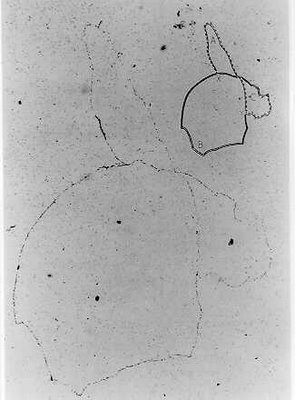
Replication mechanisms of the bacterial chromosome.

A timeless experiment in cell biology by an Australian scientist. FIGURE 5-2 from here. Autoradiograph of intact replicating chromosome of E coli. Bacteria were radioactively labeled with tritiated thymidine for approximately two generations and were lysed gently. Bacterial DNA was then examined by autoradiography. Insert shows replicating bacterial chromosome in diagrammatic form. The chromosome is circular, and two forks (X and Y) are present in replicating structure. Bar, 100 µm. From Cairns, J.P.: Cold Spring Harbor Symposia on Quantitative Biology 28:44, 1963
Schaecter et al. Microbe Chapters 8, 10 are very useful reading on this topic.
There is also the Baron free online textbook.
The chromosome of bacteria is a structure whose organisation and means of accurate and reliable duplication and distribution to daughter cells has been shaped by billions of years of natural evolution. As time passes we biological scientists are getting to understand much more of what this intense natural selection has achieved in terms of functional design.
The major points made in the teaching session were:
- The structure of a DNA molecule including the backbone and pairing of bases.
- How DNA and RNA differ.
- How a DNA molecule is replicated and what constraints the structure puts on the replication process
- A description of the proteins, including their function, that make up the replication machinery.
- How all of the steps in replication are coordinated.
Study questions to reinforce and extend points made in the teaching session:
- How are topoisomerases different to nucleases?
- What is an oriC, what are its features and what takes place there?
- What is a DNA editing activity, how does it work and why is it needed?

8 Comments:
1)How are topoisomerases different to nucleases?
The main difference between topoisomerases and nucleases is topoisomerase BIND and cut DNA then SEAL it back but nucleases only cut DNA. They DO NOT become attached and reseal DNA back.
What is an oriC, what are its features and what takes place there?
oriC stands for origin of chromosome replication. It is a specific chromosomal site where replication of chromosome begins. Replication is bidirectional, starts at oriC and ends at terminal C (terC). OriC consists of AT rich region where there are only 2 hydrogen bonding (A=T), allow the double stranded DNA easier to break open to start replication. It also has DnaA recognition sites allow the positive regulator of initiation, protein DnaA to bind and hence initiate the replication process. After DNA replication has been intitiated, origin regions specifically and transiently associate with the cell membrane leading to a model whereby membrane attachment directs separation of daughter chromosomes (the replicon model).(last sentence from Baron free online textbook)
Good to srr you actively following up on questions fam. Yes Youve said the important things about oriC.
What are the positions of oriC and ter on the chromosome? What sites and genes are near them?
What is a replichore?
a replichore is unit of replication;string of DNA where replication starts and continues to finish.
OriC and ter localise on the chromosome of opposite direction. OriC located at 84 min on the E. coli chromosome map.
I'm not sure of what genes and site near them but i found this: scattered throughout oriC are 11 GATC palindromic sequences involve in a phenomenon called sequestration to prevent the origin from participating in initiation for some time.
Fan you are doing good work. ori and ter are almost exactly opposite so bothe replichores are the same length.
Dear microbe pundit,
hi, refering lecture on this monday, i don't really know about the amplification of signal part. is it becoz of the unknown compound very little so that in order to detect it , we 'll sort of amplifying the compound? for the example u give , how to really turn one copy of gene to thousand copies of RNA? For another eg using Ts mutants, is it a tool to detect the product? and how it can be link to amplification of signal??
Thanks a lot!
Re amplification
I used the word amplification to refer to the general concept relating signal detection and its ease of measurement. If the positive signal is very small or weak, there is a need to amplify it.
A single cell is one example of a weak signal but a bacterial colony is much larger. You can amplify single cell signals by allowing them to grow to a visible colony on agar.
Other amplications work in very different ways. To amplify antibody binding reactions, chemical coupling of antibody molecules to an enzyme, like alkaline phoshatase is part of the amplification.
As far as Ts mutants I don't see any connection or relevance to this concept.
As far as very new RNA based amplification methods , don't worry about details (unless you really want to).
The point I was trying to make is that there are many alternative and innovative ways to do aplify biologically relevant signals, some yet to be invented, I'm sure.
Good question to ask though.
Thanks
The Pundit
Anyone got any suggestions on how you would design an experiment to test if a compound is mutagenic?
Post a Comment
<< Home