Friday, August 03, 2007

New biocontrol method to manage mycotoxins in Africa

NIGERIA: New hope for improved food safety in sub-Saharan Africa
01.aug.07 Via Agnet
Cross posting
Consultative Group on International Agricultural Research
Ranajit Bandyopadhyay


Scientists at the International Institute of Tropical Agriculture (IITA) have developed a safe and effective method for biological control of aflatoxins. These are toxic chemicals of fungal origin, which contaminate maize and other major food crops, posing a chronic threat to human health in sub-Saharan Africa.
With the new method, strains of the fungi that produce aflatoxin are overwhelmed through the introduction of related but entirely harmless strains. These were identified and tested through several years of meticulous research supported by the German Agency for Technical Cooperation (GTZ) and carried out in collaboration with the Agriculture Research Service of the US Department of Agriculture, University of Arizona in the USA, University of Bonn in Germany and University of Ibadan in Nigeria.

Researchers found that inoculum containing the beneficial strains can be produced most efficiently on sorghum grain, resulting in a dry formulation, which can then be broadcast on moist soil in farmers’ maize fields. Laboratory and field tests have demonstrated that the harmless strains spread quickly from the soil to maize plants, where they reduce aflatoxin-producing strains on maize grain by 91 to almost 100 percent. A single application is sufficient to control the problem in the crop treated, though a few additional applications may be required to achieve long-term control in farmers´ fields. Large-scale testing of the new method is currently under way in Nigeria.

An insidious threat
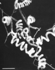
Aflatoxins are produced by various fungi, such as Apergillus flavus and A. parasiticus, which grow as molds on staple grains and root crops both before harvest and in storage. Contamination of maize causes particular concern, because it is sub-Saharan Africa’s most important cereal.
The toxins are especially damaging to children. Continuous exposure has been shown to stunt growth and even contribute to infant mortality when coinciding with kwashiorkor, a form of malnutrition caused by dietary deficiency of protein and other nutrients. The insidious combination of impaired development and undernourishment accounts for about half of the 4.5 million deaths of children under the age of 5 occurring annually in sub-Saharan Africa.
Aflatoxins are also believed to affect the human immune system, making people more vulnerable to infectious diseases, such as malaria and HIV/AIDS. In addition, the toxins are linked to liver disorders and can act in synergy with the Hepatitis B virus to cause hepatocellular carcinoma. This is the most common cancer in sub-Saharan Africa, accounting for as many as 10 percent of adult male deaths in parts of West Africa.
Aflatoxins further damage the well-being of Africa’s rural families by limiting exports of maize and groundnut in particular. Grain-importing countries maintain high food quality standards, with especially strict controls on aflatoxin content. African food and feed products showing levels of contamination above the acceptable limits cannot penetrate major grain markets, resulting in significant loss of agricultural income.
Except when people die of acute poisoning, as happened in Kenya during 2004-2006, aflatoxins seldom receive adequate attention in the region, even though they clearly have serious consequences and are quite widespread. In Benin and Togo, for example, researchers found that aflatoxin levels are about five times the safe limit of 20 parts per billion in up to 30 percent of household grain stores. According to other results from a study carried out in those countries and Nigeria, 99 percent of blood samples collected randomly from children contained aflatoxins.

In pursuit of a biocontrol strategy
In an effort to reduce aflatoxin contamination, researchers at IITA and elsewhere have deployed various methods, involving, for example, modifications in grain drying, storage and food preparation practices. To complement strategies that have already proved effective, scientists have also actively pursued in recent years the option of biological control, building on the Institute’s long and extraordinary record of success in using this approach to combat major pests such as the mango and cassava mealybugs, cassava green mite, desert locust and banana nematodes and weevils.
The biocontrol strategy that appears to be effective against aflatoxin employs a mechanism that researchers refer to as “competitive exclusion.” This is made possible by the presence in nature A. flavus populations, not only of “toxigenic” strains, which produce copious amounts of aflatoxin, but also “atoxigenic” strains, which lack this capacity. In order for the strategy to work, researchers must identify and successfully introduce harmless strains that show a large competitive advantage over the dangerous ones.
In the resulting biological struggle, explains IITA plant pathologist Ranajit Bandyopadhyay, “the good strains of A. flavus virtually eliminate their highly toxic relatives and ensure that the ‘bad guys’ cannot re-emerge.”
Such a strategy has proved effective for controlling aflatoxin on cottonseed, groundnut and maize in the USA and on groundnut in Australia.
The challenge for scientists at IITA was to identify entirely safe atoxigenic strains of A. flavus that are indigenous to Africa and serve effectively as biocontrol agents. For this purpose, they first collected more than 4,200 samples of the fungus from different ecological zones of Nigeria. In these they identified 2,127 distinct strains, of which 1,000 proved to be atoxigenic. Only 26, though, were selected for further testing.
This involved an extremely laborious series of procedures, one of them, for example, involving.more than 30,000 crosses between different strains. The purpose was to group the selected strains according to “vegetative compatibility” and make sure they belong to groups consisting only of atoxigenic strains that cannot cross with toxigenic strains in nature. This procedure ensured that release of the biocontrol agents in the field would be absolutely safe, leading to a drastic reduction, rather than an inadvertent boost, in aflatoxin levels.

Proven effectiveness
From the 14 unique atoxigenic groups identified, one strain each from eight groups was chosen for further evaluation for ability to compete with toxigenic strains. When maize grains were inoculated in the laboratory and field with both a highly toxigenic A. flavus strain as well as atoxigenic strains, one of the latter reduced aflatoxin levels by 91 percent and two others by nearly100 percent.
In experimental field plots, various atoxigenic strains proved capable of spreading quickly from the soil on which they were released to maize plants. One especially promising strain was found on nearly 99 percent of the maize grains analyzed.
The eight promising strains were further screened for their ability to get established rapidly after release, continue spreading and survive in plant debris. Strains showing these abilities can achieve effective biocontrol of aflatoxin-producing strains over multiple years, with only a few additional applications after initial release.
Having identified several excellent candidate strains to serve as agents of biocontrol, IITA researchers are further testing these in large-scale trials at various locations in Nigeria. To permit wide release of these biocontrol agents in the fields, researchers have developed safe, efficient and effective methods for producing and applying inoculum. IITA now seeks further support to disseminate the biocontrol technology as part of a “basket” of simple aflatoxin management practices that can reduce aflatoxin levels in Africa’s food and improve the health of its people.

Labels: , ,

Tuesday, July 24, 2007

Lead for drug discovery based on fundamental bacterial genetics.

New Way to Target and Kill Antibiotic-Resistant Bacteria Found

Item Courtesy Physorg.com
First Published: July 9 2007, 17:20 EST


Antibiotic resistance propagates in bacteria by moving DNA strands containing the resistance genes to neighboring cells. An enzyme called relaxase is essential for this process. Bisphosphonates, already approved to treat bone loss, have now been shown to potently disrupt the relaxase function. Some bisphosphonates prevent the transfer of antibiotic resistance genes and selectively kill bacterial cells that harbor resistance. Credit: Scott Lujan, University of North Carolina at Chapel Hill


The team discovered a key weakness in the enzyme that helps “fertile” bacteria swap genes for drug resistance. Drugs called bisphosphonates, widely prescribed for bone loss, block this enzyme and prevent bacteria from spreading antibiotic resistance genes, the research shows. Interfering with the enzyme has the added effect of annihilating antibiotic-resistant bacteria in laboratory cultures. Animal studies of the drugs are now underway.

“Our discoveries may lead to the ability to selectively kill antibiotic-resistant bacteria in patients, and to halt the spread of resistance in clinical settings,” said Matt Redinbo, Ph.D., senior study author and professor of chemistry, biochemistry and biophysics at UNC-Chapel Hill.

The study appears online the week of July 9, 2007, in the Proceedings of the National Academy of Sciences. Funding was provided by the National Institutes of Health.

The study provides a new weapon in the battle against antibiotic-resistant bacteria, which represent a serious public health problem. In the last decade, almost every type of bacteria has become more resistant to antibiotic treatment. These bugs cause deadly infections that are difficult to treat and expensive to cure.

Every time someone takes an antibiotic, the drug kills the weakest bacteria in the bloodstream. Any bug that has a protective mutation against the antibiotic survives. These drug-resistant microbes quickly accumulate useful mutations and share them with other bacteria through conjugation – the microbe equivalent of mating.

Conjugation starts when two bacteria smoosh their membranes together. After each opens a hole in their membrane, one squirts a single strand of DNA to the other. Then the two go on their merry way, one with new genes for traits such as drug resistance. Many highly-drug resistant bacteria rely on an enzyme, called DNA relaxase, to obtain and pass on their resistance genes. A mutation that provides antibiotic resistance can sweep through a colony as quickly as the latest YouTube hit.

The researchers analyzed relaxase because it plays a crucial role in conjugation. The enzyme starts and stops the movement of DNA between bacteria. “Relaxase is the gatekeeper, and it is also the Achilles’ heel of the resistance process,” Redinbo said.

Led by graduate student Scott Lujan, the team suspected they could block relaxase by searching for vulnerability in a three-dimensional picture of the relaxase protein. Lujan, a biochemistry graduate student in the School of Medicine, confirmed the hunch using x-ray crystallography, which creates nanoscale structural images of the enzyme.

The researchers predicted that the enzyme’s weak link is the spot where it handles DNA. Relaxase must juggle two phosphate-rich DNA strands at the same time. The team suspected a chemical decoy – a phosphate ion – could plug this dual DNA binding site. Redinbo, who has a background in cancer and other disease-related research, realized that bisphosphonates were the right-size decoy.

There are several bisphosphonates on the market; two proved effective. The drugs, called clodronate and etidronate, steal the DNA binding site, preventing relaxase from handling DNA. This wreaks havoc inside E. coli bacteria that are preparing to transfer their genes, the researchers found. Exactly how bisphosphonates destroy each bacterium is still unknown, Redinbo said, but the drugs are potent, wiping out any E. coli carrying relaxase. “That it killed bacteria was a surprise,” he said. By targeting these bacteria, the drugs act like birth control and prevent antibiotic resistance from spreading.


see also National Review of Medicine

(Note J. Chan is considering this topic for her bioproduct proposal)

Labels: ,

Saturday, June 16, 2007

Energy and gold among the SliMEs

Clean fuel research deal agreed by BP and Synthetic Genomics
15.jun.07
Biofuel Review
Synthetic Genomics Inc., a privately-held company dedicated to commercializing synthetic genomic processes and naturally occurring processes for alternative energy solutions, has announced a significant, long-term research and development deal with BP.
The deal between BP and Synthetic Genomics is centered on developing biological conversion processes for subsurface hydrocarbons that could lead to cleaner energy production and improved recovery rates. As part of the agreement, BP has also made an equity investment in Synthetic Genomics.
Microbes are key components in sustaining and maintaining life on Earth, and genomics is leading to an enhanced understanding of these organisms. In the first phase of the BP/Synthetic Genomics program, the research will focus on gaining a better understanding of microbial communities in various hydrocarbon formations such as oil, natural gas, coal and shale. Synthetic Genomics, which was founded by genome pioneer J Craig Venter, Ph.D., will use its expertise in environmental DNA sequencing and microbial cell culturing to produce the first comprehensive genomic study of microbial populations living in these environments. Once the basic science research phases are complete, BP and Synthetic Genomics will seek to jointly commercialize the technologies developed.
“We believe that one of the most promising solutions to producing cleaner fuels will be found through genomic-driven advances,” said Dr. Venter. “Through our research collaboration with BP, we will achieve a new and better understanding of the subsurface hydrocarbon bioconversion process which we are confident will yield substantial cleaner energy sources.”
The overall goal of Synthetic Genomics is to discover and/or design new genomes that will code for new types of cells with desired properties for bioenergy or specific chemical production. In this project, Synthetic Genomics scientists hope to better understand hydrocarbon metabolism by sequencing the genomes and culturing the cells of the naturally occurring microbes that thrive in subsurface hydrocarbons.
Tony Meggs, Group Vice President of Technology at BP, adds: “This collaboration is an exciting development that could lead to an unprecedented understanding of the microbial activity in the subsurface; and eventually to the development of more environmentally-friendly and efficient energy production and recovery techniques.”

Labels: , , ,

Tuesday, October 31, 2006

Q and A on BACs and Metagenomics. Why large inserts?

Question from student (edited):

I am reviewing the Soil Metagenome paper that we reviewed in class (Rondon et al., 2000, Appl. Environ. Microbiol. 66: 2541-2547), and have a written note claiming that there are two important reasons to clone large DNA fragments into the BAC vectors. I can only think of one reason, which is that many soil microbes produce important bioactive compounds, and genes required for production along with regulatory genes are often clustered in one continuous segment on the chromosome called an operon. A large fragment of DNA inserted into BAC plasmid would increase the likelihood that the entire operon included. Is this correct, and is there another reason why having large DNA fragments in the BAC plasmids is important?

Answer:

Yes, you are correct, and yes, there is another reason. Large inserts mean that each clone (Escherichia coli clone line) is likely to contain genes encoding for multiple activities that may be of interest. This means that fewer clones need to be maintained than if the inserts were smaller, i.e., the same amount of metagenomic information is maintained in fewer clones (or more metagenomic information can be maintain in any given number of clone lines). Using screening assays for different activities, the same amount of metagenomic material can therefore be interrogated for the presence of specific activities while manipulating fewer cultures.

Labels: , , , ,

Monday, October 30, 2006

Discovering novel antifungal agents with antimitotic activity from diverse soil and water sources.

C W S Fen, Melbourne

Abstract
In response to the side effects and increasing amount of resistance met by antifungal drugs targeting the ergosterol pathway in fungi, we propose to bring antimitotic antifungal agents to a reformation, enlisting the use of high throughput screening to discover novel microtubule-inhibiting compounds produced by fungi from a diverse range of soil and water sources. We intend to screen large amounts of secondary metabolites produced by these fungi against 10 specifically chosen common infection-causing fungi species. After narrowing down the candidate compounds to a few lead compounds, these will be tested for fungal-specific antimitotic activity in vitro with fungal microtubule dimers and in vivo against the 10 fungi species utilizing GFP-labelling technology. Toxicity to human cells will be assessed as well before clinical trials can be proposed. Further experiments are necessary to determine optimum dosage and method of delivery of the drug, and further research into potential anticancer drug applications may yield additional commercial benefit from investment in this initiative.

Introduction
Imagine if a harmless scab that you thought would just go away became the cause of your hospitalisation. Or the joy and relief of being discharged after an operation only to find yourself back in bed being treated for a post-surgical fungal infection. Fungal infections have a significant impact on the quality of life of affected individuals despite little attention dedicated to it when compared to other life-threatening diseases. However, most predominantly, it is patients who have undergone surgery, are immunocompromised, have undergone chemotherapy or excessive antibiotic doses who are severely affected and whose life may well be in danger if the infection is not addressed immediately (White et al., 1998). In view of this, biotechnological innovations and advances over the past few years can be put to good use in fuelling the initiative of discovering new or improved anti-fungal compounds which targets fungi specifically while sparing human cells.

In light of this, we propose to bring fungal-specific antimitotic drugs to a reformation, achievable by exploiting the fungal cells’ dependence on microtubule function to proliferate, and thus prevent the progression of infection. From a marketability standpoint, the diverse range of pharmaceuticals that target the ergosterol pathway in fungi are saturating the market, resulting in resistance among different drugs targeting this same biochemical pathway (White et al., 1998). Therefore the guaranteed effectiveness of this new drug minus the side effects of its competitors (which are the main source of concern), coupled with increased efficacy, is highly promising in guaranteeing a high return in investment. Accordingly, we intend to perform high-throughput screening of soil and water samples from a diverse range of sources to screen for fungi that have the ability to produce such a compound. Following that, we will run carefully designed assays to engineer additional broad-spectrum activity of the said compound. Miracles are more likely to happen if we make them and we intend on doing just that.

PROPOSAL:

The Scientific Basis

Microtubules are essential to all eukaryotic cells; in essence, fungal cell shape, transport, motility and most importantly for this proposal, cell division, rely wholly on the proper function of this critical organelle. The fundamental property of microtubules that we look to exploit in a biotechnological sense is its non-equilibrium behaviour, better known as microtubule dynamic instability, which determines the shortening or lengthening of microtubules to facilitate them in performing the functions listed (Nogales 2001). With this in mind, we analysed fungal cell characteristics to determine a logical strategy of attack.

Many secondary metabolites produced by fungi have potent pharmacological uses through exploiting its initial intended function. An obvious example of this is Penicillin produced by the mould Penicillium notatum a commonly used antibiotic for treating bacterial infections and diseases. However, this same species of mould from which penicillin is derived also produces another compound, Griseofulvin, synthesised by a close relative, Penicillium griseofulvum, which prevents microtubule formation in fungal cells during mitosis. Although griseofulvin requires a longer duration of action and exhibits less efficacy than other commonly used antifungals, nevertheless, it boasts minimal side effects and a currently still unexplored biochemical mechanism as to why it targets fungal cells specifically, partially sparing human cells and therefore resulting in milder side effects (Jimenez et al, 1990). Logically assuming that it is the specific action or higher affinity of the drug for fungi microtubules that results in the milder side effects, we propose to further expand on the merit of this particular target protein of the drug and search for novel compounds which utilises a similar mechanism of attack to halt the growth of fungi, making it a potent fungistatic agent. Subsequently (not covered by this proposal), chemical modification or further subjection of the fungi species of interest to selective chemicals or mutagenic agents can be run to develop a compound that will bind irreversibly to the microtubules and thus effectively kill off the target fungal cells (Refer to Figure 1 and Figure 2 below).

Figure 1: Two possible models of microtubule nucleation. (a) End-on model of nucleation; (b) Lateral interaction of γ-Tubulin protofilament with αβ-Tubulin dimers. (Modified from Nogales, 2001). Irreversible binding of the antifungal compound (in red – whether to the protofilament as whole or to individual dimers) can prevent prolongation of αβ-Tubulin protofilament or interaction between filaments, disrupting microtubule instability and therefore mitosis.


Figure 2: Ribbon diagram of molecular structure of α- and β-tubulin dimer of microtubules. A compound that can bind irreversibly to a point on this most basic unit of microtubule structure at e.g. the taxol binding site (circled in red) can trap these tubulin dimers and cause stabilization of microtubules, preventing mitosis (Revised from Nogales, 2001).


The Source

We plan to run high throughput screening of a range of soil and water samples obtained from diverse geographical locations around the world to isolate as many species of fungal cells as possible, with a more bias focus on mycoparasites, as toxic compounds produced by fungi against other fungi will be more specific and therefore more effective. Then, we will narrow down the candidates of these fungal species to those that may harbour antimitotic activities.

By diluting the soil sample in water and partially diluting the water samples, multiple cultures (100 odd petri dishes) of fungi are obtained using growth media that will stimulate fungal growth, e.g. YES media (Torres et al., 1987). After this primary culture, each fungal colony will be extracted and cultured at diluted concentrations in zone-based bioassays overlaid with a layer of one of 10 designated common infection-causing fungi, i.e. the Tinea spp., Candida spp., Aspergillus sp., Cryptococcus spp., Fusarium spp., etc. (Mays et al, 2006). It was shown by Panda et al., 2005, after a few weeks, any fungal colony whose surrounding cells are trapped in interphase and highly mononuclear exhibits antimitotic activity. These colonies are isolated and the compound produced by each colony is extracted and purified by chemical means. These compounds are then subjected to high throughput screening to test for antimitotic activity.

High Throughput Screening

The screen we have in mind is designed to be a fungal-specific antimitotic screen. Exploiting the success of genomic sequence technology and the completion of function determination of thousands of genes, we will search for genes encoding microtubule function in the 10 species of common infection-causing fungi using the available genome libraries online. By ensuring that the 10 chosen species of fungi have been studied intensively enough to for us to genetically manipulate the microtubule and its associated proteins’ genes, hypersensitive mutants susceptible to antimitotic drugs will then be generated and these mutant fungi will be used to screen for compounds that can inhibit their growth among the potential candidate compounds produced by the initial fungal culture. Testing will be done at increasing concentrations of antimitotic/antifungal compounds. Following positive results, the wild type strain for each of the 10 common disease causing fungi is then exposed to these 10 compounds as a control to identify any of the compounds can arrest the cell cycle of the parent strain (Lila et al, 2003). In this way, the antimitotic drug-sensitive fungi will increase the likelihood of discovering a drug that has antimitotic potential which we can improve the properties of through further screening.

Thus, narrowing down the search to one lead compound, we will proceed to test this in vitro with microtubules obtained from fungal cells. By tagging the fungal tubulin dimer proteins with GFP tags, we will then monitor the action of the compound on the polymerization of individual tubulin entities with increasing lead compound concentration.

From in vitro studies, we will extrapolate success of the compound in inhibiting microtubule function to in vivo testing by a reporter system where cloning technology is used to introduce fluorescence into microtubule dimers by incorporating the GFP gene into the tubulin gene (cf. protein) of each of the 10 fungal species which is then transcribed into tubulin dimers that will fluoresce, thus enabling us to monitor the growth or shrinking of microtubules (mitotic spindle) during mitosis (Implication of concept by Kumagai et al., 2003 and Alberts et al., 2002). Further proof can be obtained by running flow cytometry analysis of fungal cells treated with cytotoxic concentrations of the lead compound – if there is no increase in nuclear DNA content, then there is a lack of tubulin-related activity in vitro (Lila et al, 2003).

These same set of in vitro and in vivo experiments are repeated with human tubulin and in human cells from various tissues across the body respectively to test for toxicity of the compound (Priestly and Brown, 1978). Thus, if luck it working on our side, we may be able to determine a distinct fungal specificity of the drug and determine the exact concentration of the compound which is the threshold of toxicity for human cells. Complicated but necessary further experimentation should be done in whole organism model systems such as rats, mice and other mammals to mimic possible side effects in humans before proposing the extrapolation of these results to humans in clinical trials.

Potential Problems

Microtubules, despite being in different organisms, are highly conserved organelles, especially between organisms of the same domain (Eukaryota). We are taking a significant risk in gambling with nature on the possibility that antifungal compounds produced by fungi themselves against their fungal counterparts might specifically inhibit these competitors for survival antimitotically. At the very least however, we can design these novel compounds specifically for a distinct group of fungal species (e.g. specifically one of superficial, subcutaneous, systemic or opportunistic infection-causing groups of fungi) if not for broad-spectrum activity against most of them.

As in any microbial biotechnology process, transition from a small-scale experiment to culturing of these fungi of interest in the large scale will present us with many more problems to come. However, given the appropriate equipment, competent technicians and proficient biotechnologists, this should be a challenge we can overcome.

Further Research

As part and parcel of an antifungal treatment, side effects are inevitable as human cells are very similar structurally to fungal cells, as mentioned earlier. Thus, drugs that target fungal cells will affect our cells to a certain extent as well. Therefore, although the side effects will be specifically tailored to be minimal, further chemical modification or manipulation (if possible) to obtain compounds that have near-negligible side effects will be needed following the isolation of the fungal microtubule inhibitor protein-producing fungi..

In addition to that, also on a pharmacological basis, we need to take into account the stability of the compounds (determining optimal pH), scaling up of production and the efficiency of cell growth of the cell of interest in culture. Other than that, solubility of antifungal compounds is a major concern in the pharmaceutical industry as most antifungal compounds are contrary to that requirement. Added to that oral bioavailability, potential side effects and possible allergic reactions have to be accounted for to guarantee its economic value.

Potential Future Benefits (Integration of Science and Business)

One of the key valuable gains from the production of this initial microtubule formation inhibitor compound is a better understanding of the mechanism of microtubule inhibition and prevention of mitosis. From investigating so many fungi species with antimitotic activity, we can gain an insight into the many potential ways the microtubule can be inhibited and by manipulating this knowledge as well as other biotechnological screening and assay methods to slightly modify the compound, we could extrapolate the research to involve discovery of similar compounds from fungi which will specifically inhibit microtubule formation of tumour cells and thus secure a spin-off opportunity to produce a potential anti-cancer drug. Much research efforts and funds have been poured into a similar initiative involving Griseofulvin (Panda et al., 2005). Thus, this presents a further bonus for investing in this research.

Conclusion

In conclusion, this initiative of discovering novel antimitotic antifungal drugs presents many commercial, scientific and medical benefits to every player: investors, consumers and the biotechnologists. Besides solving a demanding medical problem and gaining much knowledge about fungi and its infection-causing mechanisms, there is much to gain from investment in the long run, with spin-off opportunities that are becoming a reality in the anticancer drug industry. “Fortune favours the prepared mind - Louis Pasteur.” This statement could not hold truer in this fascinating hybrid between business and science.

References
1. Lila, T., Renau, T.E., Wilson, L., Philips, J., Natsoulis, G., Cope, M.J., Watkins, W.J. and Buysse, J. 2003. Molecular Basis for Fungal Selectivity of Novel Antimitotic Compounds. Antimicrobial Agents and Chemotherapy. 47: 2273-2282.

2. Nogales, E. 2001. Structural Insights into Microtubule Function. Annual Review of Biophysics and Biomolecular Structure. 30:397-420.

3. Kumagai, F., Nagata, T., Yahara, N., Moriyama, Y., Horio, T., Naoi, K., Hashimoto, T., Murata, T. and Hasezawa, S. 2003. Gamma-tubulin distribution during cortical microtubule reorganization at the M/G1 interface in tobacco BY-2 cells. European Journal of Cell Biology. 82:43-51.

4. Alberts, B., Johnson, A., Lewis, J., Raff, M., Roberts, K. and Walter, P. 2002. Molecular Biology of The Cell, 4th edition. Garland Science, New York, USA.

5. White, T.C., Marr, K.A. and Bowden, R.A. 1998. Clinical, Cellular, and Molecular Factors That Contribute to Antifungal Drug Resistance. Clinical Microbiology Review. 11: 382-402.

6. Torres, M., Canela, R., Riba, M. and Sanchis, V. 1987. Production of patulin and griseofulvin by a strain of Penicillium griseofulvum in three different media. Mycopathologia. 99: 85-89.

7. Mays, S.R., Bogle, M.A. and Bodey, G.P. 2006. Cutaneous Fungal Infections in the Oncology Patient: Recognition and Management. American Journal of Clinical Dermatology. 7: 31-43.

8. Priestly, G.C. and Brown, J.C. 1978. Effects of griseofulvin on the morphology, growth and metabolism of fibroblasts in culture. British Journal of Dermatology. 99: 245.

Labels: ,

Step up: Bacterial Lipase to Substitute Pancreatic Lipase For Enzyme Therapy

D. G. Sani, Melbourne

Summary
Microbial lipases have been more commonly used than lipases derived from plants and animals due to higher stability, rapid production, higher yields and more convenient manipulations of microorganisms. For example, lipase from Mucor miehei is used in cheese production, Candida antartica in surfactants, Serratia marcescens lipase in drugs (diltiazem). Medical area is another promising area where lipase can be used as digestive aids. To date, only pancreatic enzyme (lipase) therapy is used to treat fat malabsorption in Cystic Fibrosis and pancreatitis patients. However, pancreatic lipase is susceptible to low pH (acidic gastric environment) and protease which can render the lipase inactive. Therefore, it is proposed that an alternative bacterial lipase might have the ability to retain its activity under acidic condition and also be protease tolerant. Screenings for this novel bacterial lipase would be done from bacterial populations that are known to survive in the gastric environment. Moreover, different assays would be done to observe the lipase production, activity, and specificity under certain conditions.


A. INTRODUCTION

Bacterial lipases have been recognized since nearly 100 years ago in lipase-producing bacteria such as Bacillus pyocyaneus (today is named Pseudomonas aeruginosa), Staphylococcus pyogenes (S. aureus), B. fluorescens (P. fluorescens) [1]. For a lipase to be defined as a true lipase, it must exhibit interfacial activation where a presence of triglyceride should rapidly increase its activity; it should also contain a loop allowing substrate entry to the active site. However, these two criteria are unsuitable for some exceptions where the lipases have the loop but do not exhibit interfacial activation. Hence, a simpler definition of lipases is carboxylesterases that catalyse the hydrolysis and synthesis of long-chain acylglycerols [1]. The prospect for industrial enzymes (technical, food, and animal feed enzyme) is very promising with an estimated increase to $2.4 billion in 2009. Technical enzyme such as in detergents, pulp production, and medical treatment is predicted to have the highest share at 52% [2].


Global Enzyme Markets Based on Application Sectors, 2002-2009
($ Millions)

Source: BCC research [2]


Application of lipase in industrial enzyme production has covered a wide area: flavour development in food technology; as oil removal in detergents; textile making; personal care products e.g. cosmetics; digestive aids for medical treatment [3]. As digestive aids, pancreatic lipase has been widely used in the enzyme therapy to treat lipid malabsorption e.g. in cystic fibrosis and pancreatitis patients. However, there are some barriers with the pancreatic enzyme therapy that prevent its maximal efficacy [3]. Hence, the need of finding an alternative lipase from microorganisms has been raised. Microbial enzymes have several advantages than animals or plants enzymes due to the stability, high yields and rapid production from microbes, and variety in catalytic activity [3]. This paper will firstly describe the background and target market of enzyme therapy for lipase, followed by proposal of methods to isolate lipase-producing bacteria and assays to obtain the acid and protease tolerant lipase.


B. BACKGROUND AND TARGET MARKET
Cystic Fibrosis (CF) is an autosomal genetic disorder affecting mostly Caucasian populations with approximately one in every 3500 newborns is born with CF each year in the United States alone [4]. Being a multi-system disease, CF affects many organs: lungs, pancreas, liver, bones (osteoporosis) [5]. The altered gene in CF disrupts the function of salt transports in organs leading to an excess production of thick, sticky mucus that blocks the ducts in these organs mainly the lungs and the pancreas. Patients with affected pancreas tend to have digestion and growth problems due to pancreatic insufficiency where there is a lack of pancreatic enzyme produced by the body [4,5]. This condition leads to lipid malabsorption because the body cannot break down the essential fatty acid (EFA) molecules coming from external nutrients [6]. Poor nutritional status due to EFA deficiency is correlated with weight and height retardation in children with CF. Moreover, a research by CDC in the US has shown a correlation between nutritional status and the lung function showing increase in body weight in CF patients (measured by BMI) is accompanied by increase in the lung function [4].

Many treatments have been developed to treat the malnutrition in CF patients and improve their life expectancy. To date, pancreatic enzyme (lipase) therapy from porcine origin has been the most widely used since 1930s with about 90% of the patients taking pancreatic enzyme supplements in 2004 [4, 7]. However, the efficiency of this treatment has not reached maximal yet. The pancreatic lipase is found to be denatured by gastric acids before reaching the small intestine where the FA hydrolysis and absorption mainly occur. Although coating the enzyme with an acid resistant (enteric) coating appears to improve the delivery of the enzyme into the small intestine, the low intestinal pH in CF patients after meal (pH less than 4) would still irreversibly denature the pancreatic lipase [7].

Another barrier for the existing pancreatic enzyme therapy is that pancreatic lipase is also denatured by proteases especially chymotrypsin in the intestinal lumen [7]. Hence, in search of ways to improve the enzyme therapy, the aim is to find an alternative lipase that would be acid resistant, protease resistant and would still possess the pancreatic lipase activity.


C. SCIENTIFIC BACKGROUND FOR BACTERIA ISOLATION
In order to find a lipase which shares features with pancreatic lipase, one must know the characteristics of pancreatic lipase. It is known that pancreatic lipase hydrolyses long-chain triacylglyceride (TG) into FAs to be readily absorbed in the small intestine at the optimal pH of 8 [6]. When the pH falls below six the pancreatic lipase is rendered inactive and if the pH less than 4.5, it is irreversibly inactivated; the later is the case in CF patients causing denaturation of the pancreatic lipase in the small intestine during the enzyme treatment [6, 7]. Hence, the alternative lipase would have to be able to survive under acidic environment (pH less than 4). Moreover, knowledge of the active site structures of pancreatic lipase can be a valuable information in finding a new lipase. The active site residues of pancreatic lipase are serine (Ser 152), aspartate (Asp 176), and histidine (His 263) [8]. These are found to be conserved in bacterial lipases where the active site has a nucleophilic residue (Ser, Cys or Asp), a catalytic triad residue (Asp or Glu), and a histidine [1].

Acid resistant lipase could most probably be obtained from bacteria living in acidic environment. A study shows that Salmonella, a gastrointestinal pathogen, produces proteins: RpoS, PhoP, and Fur that help them survive under the harsh gastric environment. RpoS and Fur are responsible for survival against organic (weak) acid stress, while PhoP and RpoS act against inorganic acid (low pH) stress [9]. Therefore, bacteria that produce RpoS, PhoP and Fur would presumably be able to survive under low pH in the gastric environment. A database search from the genbank of NCBI returns 33 bacterial strains that have RpoS, PhoP, and Fur proteins in their genome. Most of these strains are from Salmonella, Pseudomonas, E. coli, Yersinia, Shewanella, and Shigella [10].

Out of these bacterial populations, previous studies have shown that some Pseudomonas strains produce true lipase while both E. coli and Salmonella typhimurium produce esterases [1]. Bacteria surviving from the gastric environment would most probably be present in faeces. Hence, novel lipase-producing bacteria can be screened from faecal specimens and looking specifically for Yersinia, and Shigella as no lipases have been identified from these genera before. These specific strains can be isolated from faecal specimens using selective media. For example, MacConkey agar (MAC) is used to isolate lactose-fermenting, gram negative enteric bacteria while xylose lysine desoxycholate (XLD) agar is specific for isolation of Salmonella and Shigella [11]. Yersinia can be isolated on DYS medium containing peptone, ox-bile, arginine, lysine, arabinose, casein where Yersinia will ferment arabinose and appear as bright red colonies [12].

Another consideration in searching for a new lipase is the specificity of the lipase. Since the aim is to screen for a bacterial lipase that mimics pancreatic lipase, the bacterial lipase should be able to hydrolyse long-chain TGs- the substrate for pancreatic lipase [6]. Although the term ‘long-chain’ TG is not strictly defined, in general, glycerolesters with an acyl chain length more than ten carbon atoms can be considered as lipase substrates e.g. trioleoylglycerol. On the other hand, esterase substrates would normally have an acyl chain length less than 10 carbon atoms e.g. tributyrylglycerol. Most lipases, however, are capable of hydrolysing esterase substrates in addition of the lipase substrates [1]. Hence, screening for the alternative lipase would differentiate between lipase and esterase specificities.

D. ASSAYS
Assays that would be used for screening of an alternative bacterial lipase are assays for lipase production, lipase purification, activity and specificity, pH effects on activity (low pH), and tolerance on protease (chymotrypsin).

Identification of lipase-producing bacteria from the formerly isolated strains can be done by a plate assay where the agar medium contains triolein as the lipase substrate. Lipase production on triolein plates is indicated by orange-red fluorescence of the colonies at 350 nm UV lamp [1]. The lipase-producing colonies can then be purified by inoculating a single colony onto agar plates containing essential compounds for growth. Then the strains would be grown in minimal media containing some essential nutrients for growth and enhance extracellular lipase production. It has been studied that exolipase production in Serratia marcescens can be enhanced by addition of certain polysaccharides as shown in the table below [13].

Table from Winkler, U., Stuckmann, M. (1979). Glycogen, hyaluronate, and some other polysaccharides greatly enhance the formation of exolipase by Serratia
marcescens. J Bacteriol 138: 663-670. Not displayed.

Table Source: Winkler [13].

It can be seen that laminarin exhibits the highest exolipase inducing ability followed by glycogen and pectin B, then hyaluronate. Moreover, the exoprotease production should be kept to minimum so that it would not degrade other essential proteins. Amongst these exolipase enhancers, the most suitable polysaccharide would be pectin B since it has high inducing ability with less exoprotease activity produced compared to laminarin and glycogen. Furthermore, due to interfacial activation of most lipases, addition of long-chain triglyceride such as olive oil might enhance the activity. Hence, pectin B and olive oil can be included in the medium as exolipase enhancers.

There are different assays to test lipase activity in the supernatants namely: the photometric assay and titration method [1]. The photometric assay is done using p-nitrophenylpalmitate (pNPP) as the substrate which is mixed with the supernatant [13]. Lipase activity is then monitored by detecting the hydrolysis of various p-nitrophenolesters of FAs (greater than 10 carbon atoms) into p-nitrophenol at 410 nm. Different p-nitrophenolesters would be separately included in the reaction mixture to test for lipase specificity [14]. The titration method measures lipase activity by recording the amount of NaOH used to maintain pH 8 as the fatty acid is liberated after addition of lipase solution into the supernatants [15].

Assay to screen for acid tolerant lipase can be performed by setting up a reaction mixture mimicking the gastric environment where the pH can be as low as 2.5 to 4. The synthetic gastric fluid has been widely used by mixing pepsin, lysozyme, and bile. Although the synthetic gastric fluid is more convenient to prepare, it has been demonstrated that certain bacteria are more sensitive in an ex vivo porcine gastric fluid than in the synthetic gastric fluid at a same pH of 2.5. This suggests that there are some components which are present only in the porcine stomach and can interfere with bacterial survival [9]. Hence, a more reliable assay for acid tolerant lipase can be done using porcine gastric fluid collected from live pigs instead of the synthetic gastric fluid. The pH of porcine gastric fluid can be adjusted from 3.47 to 4.15 by mixing supernatants of several stomach contents with pH ranging from 1.42 to 4.44 [9]. This mixture (synthetic or porcine gastric fluid) can then be added into the bacterial supernatants and then assayed for lipase activity (photometric assay). The acid resistant lipase would retain its ability to hydrolyse p-nitrophenylesters after treatment with the gastric fluid.

Protease tolerant lipase can be assayed by adding a specific protease into the bacterial supernatants prior to the lipase activity assay. For this experiment, chymotrypsin would be used since it is found to denature pancreatic lipase in the small intestine [7]. If successful, the bacterial lipase that retains its activity would be the protease tolerant lipase. After confirming the lipase which possesses the specific characteristics, purification of the lipase can be done by chromatographic methods where the lipase is bound to beads in a column and then eluted with corresponding buffer. For instance, extracellular lipase of Pseudomonas was purified by anion-exchange chromatography and HIC [15]. The main advantage is that purified enzymes have high activity and can be re-used in other reactions.

CONCLUSION
Technical enzymes application in various biotechnology areas has shared the highest part in the global enzymes market since 2002 [2]. One of the major areas is in medical application where pancreatic lipase therapy is used as digestive aids mostly in CF patients. However, the efficacy has not reached maximal due to degradation of the lipase under acidic condition in the stomach and small intestine of these patients [7]. Hence, searching an alternative acid-tolerant lipase from bacterial origin to substitute pancreatic lipase as digestive aids would have high market returns in the future. If successful, the novel bacterial lipase would be acid and protease tolerant where it can retain its activity at pH less than 4. Moreover, purification would increase its activity implying for higher end products yields from fewer lipase mass and hence, effective production cost.

REFERENCES

1. Jaeger, K. E., Dijkstra, B. W., Reetz, M. T. (1999). Bacterial biocatalysts: molecular biology, three-dimensional structures, and biotechnological application of lipases. Annu Rev Microbiol 53: 315-51.

2. BCC report on global enzyme market. http://www.bccresearch.com/bio/BIO030D.asp. Retrieved 5/10/2006.

3. Hasan, F., et. al. (2006). Industrial applications of microbial lipases. http://www.aseanbiotechnology.info/Abstract/21019024.pdf

4. Cystic Fibrosis Foundation. (2004). Patient registry annual data report 2004.

5. Gotz, M. H. (2002). Cystic Fibrosis literature review annual report 2002.

6. Peretti, N., et. al. (2005). Mechanisms of lipid malabsorption in Cystic Fibrosis: the impact of essential fatty acids deficiency. Nutrition & Metabolism 2:11.

7. Durie, P., et. al. (1998). Uses and abuses of enzyme therapy in cystic fibrosis. J R Soc Med 91: 2-13.

8. Olivecrona, G. et al. (1994). “Medical aspects of triglyceride lipases.” In P. Woolley & S.B Petersen (Eds.), Lipases: their structure, biochemistry and application pp.316-318.

9. Bearson, S. M. D., et. al. (2006). Identification of Salmonella enterica serovar Typhimurium genes important for survival in the swine gastric environment. Applied and Environmental Microbiology 72: 2829-36.

10. NCBI web site: http://www.ncbi.nlm.nih.gov/entrez/query.fcgi?db=Genome&cmd=search&term=RpoS,+Fur,+PhoP+containing+bacteria Retrieved 29/9/2006.

11. CDC: Ch4: Identification and isolation of Shigella. http://www.cdc.gov/ncidod/dbmd/diseaseinfo/cholera/ch4.pdf#search='isolation%20of%20shigella'. Retrieved 4/10/2006.

12. Agbonlahor, D. E., et. al. (1982). Differential and selective medium for isolation of Yersinia enterocolitica from stools. J Clin Microbio 15: 599-602.

13. Winkler, U., Stuckmann, M. (1979). Glycogen, hyaluronate, and some other polysaccharides greatly enhance the formation of exolipase by Serratia
marcescens. J Bacteriol 138: 663-670.

14. Kanwar, S. S., et. al. (2006). Purification and properties of a novel extracellular thermotolerant metallolipase of Bacillus coagulans MTCC-6375 isolate. Protein Expression & Purification 46: 421- 428.

15. Kordel, M., et. al. (1991). Extracellular lipase of Pseudomonas sp. strain ATCC 21808: Purification, characterization, crystallization, and preliminary X-Ray diffraction data. J Bacteriol 173: 4836-41.

Labels: , ,

Monday, October 09, 2006

Screening for Novel Anti-cancer Bioactive Compounds Produced by Bacteria Found in Marine Sponges.

A Proposal for Novel Compound Discovery

H. A. Ward, Melbourne


Abstract
The chemical and biological diversity of the marine environment is immeasurable and is therefore an extraordinary resource for the discovery of novel anti-cancer compounds. Recent technological and methodological advances in structure elucidation, biological assays and organic synthesis have enabled the isolation and clinical evaluation of various novel anti-cancer agents from marine microorganisms. This report provides insight into the processes involved in the isolation and identification of novel marine microorganisms, the screening for novel bioactive anti-cancer compounds produced by the microorganisms, as well as the evaluation and characterization of their structure and bioactivity.


Introduction
With an estimated 7.6 million deaths reported in 2005, cancer is the second leading cause of death amongst the world’s population . Each year, several billion dollars is invested in cancer research in an attempt to understand the disease processes involved and to try to discover possible therapies to combat this debilitating disease. The potential market for immune drugs is huge and is a rapidly growing area in the biopharmaceutical arena. It is expected that there will be a US $15 billion worldwide market opportunity for therapeutic immune drugs by the year 2010 .

During the past 5 decades of research in anti-cancer drug discovery, about 100 products have been provided for clinical treatment of malignancy (Wagner-Dobler et al., 2002). Significant progress has been made in the chemotherapeutic management of hematologic malignancies, however, more than 50% of patients with tissue tumours either fail to respond or will die from the disease (Wagner-Dobler et al., 2002). Hence, the discovery of novel anti-cancer therapeutic agents remains critically important.

The tremendous biochemical diversity of marine microorganisms and their biotechnological potential, as extraordinary resources for the discovery of new anti-cancer compounds, is becoming more and more recognized by both microbiologists as well as the pharmaceutical industry (Schweder et al., 2005). Of these microorganism species, the microorganisms living in marine sponges have attracted significant attention as potential sources of bioactive compounds (Garcia Camacho et al., 2006). Because of their phenomenal potency, even very small quantities of these compounds can be of significant value in a commercial sense (Simmons et al., 2005).

Culture based techniques are inadequate for studying bacterial diversity from marine samples as many bacteria cannot be cultured using artificial laboratory conditions (Webster et al., 2001) and thus, is not accessible for detailed taxonomical and physiological characterizations. The tools of molecular biology and the phylogenetic framework, which is now available as 16S rDNA sequence alignments, allow a complementary strategy to be pursued. This is based on phylogenetic screening of marine isolates and an in-depth investigation of the biological activity and chemical diversity of selected phylogenetic groups in order to identify a new hotspot for the production of bioactive compounds (Schweder et al., 2005). Selected isolates can then be cultivated on a large scale and under a variety of cultivation conditions.
This report aims to provide an insight into the steps which are involved in screening for these novel anti-cancer bioactive compounds which are produced by marine bacteria.

Proposal
Searching for novel bioactive compounds is a multi-step procedure which begins with the selection of a suitable source to be investigated- in this case, it is a sample of sponge tissue which will contain numerous bacteria, producing bioactive compounds (Wagner-Dobler et al., 2002). The sponge tissue is collected by scuba-diving in the region where the marine sponges are located. The phylogenetic affiliation of the sponge-associated bacteria is assessed using16S rDNA analysis which is mainly based on the selective amplification of 16S rRNA gene sequences, of taxon-specific lengths, by the application of primers of conserved regions of the 16S rDNA in combination with PCR and gel electrophoresis (Schweder et al., 2005). Due to its highly conserved and variable sequence regions, the 16S rDNA sequence is used as a phylogenetic marker (Schweder et al., 2005). This method of direct isolation of DNA from the environment and cloning and sequencing of 16S rRNA genes enables identification and taxonomical affiliation of bacterial species in different natural marine habitats without the cultivation of the microbial cells.

Production of secondary metabolites is usually a strain-specific trait. Thus, typing of the isolated bacteria with a high resolution is necessary to assess the genetic diversity of the strains within a given phylogenetic group (Wagner-Dobler et al., 2002). This high resolution is obtained by using genomic fingerprint methods- in this case a RAPD (Random Amplified Polymorphic DNA) technique with arbitrary primers is used (Wagner-Dobler et al., 2002).

Additionally, phylogenetic data on microbial community composition in sponges can indicate possible nutritional requirements and physiological niches of many microbes based on information already available for known phylogenetic relatives (Webster et al., 2001). This may assist in the experimental manipulation of culture conditions to provide the correct growth environment for targeted bacteria (Webster et al., 2001).

One of the limitations associated with the construction of 16S rDNA clone libraries from total environmental DNA is that it requires the use of PCR, which precludes quantitative estimates of abundance for each organism (Webster et al., 2001). This can be overcome to some degree by the use of fluorescence in situ hybridization (FISH) probing. FISH with rRNA specific probes allows phylogenetic identification of bacteria in mixed assemblages and enables the cells to be visualized and semi-quantified (Webster et al., 2001).

The crude extracts are investigated to evaluate anti-bacterial, anti-fungal, phytotoxic or cytotoxic activity. Individual bacterial colonies are obtained by serially diluting the sample and spread-plating appropriate dilutions on agar plates containing a variety of marine media (Wagner-Dobler et al., 2002). Brine shrimp toxicity has a strong correlation with cytotoxicity and is therefore a good indicator for potential anti-cancer activity (Wagner-Dobler et al., 2002). Secondary metabolite production can only be assigned to the bacteria when synthesis has been demonstrated in cultures isolated from the host species (Webster et al., 2001).

The plates are then incubated at room temperature for up to 4 weeks. The growth of eukaryotes and protozoan grazing is prevented by the addition of the antibiotic cycloheximide (Wagner-Dobler et al., 2002). The isolated bacteria are then compared to the total community structure of the sample determined by the small subunit rDNA approach. Novel bacterial status is assigned to isolates after comparison of colony morphotype and microscopic appearance of gram-stained preparations with previously obtained isolates (Webster et al., 2001).

The strongly positive hits obtained using the serial dilution and agar diffusion tests are then screened further using tumour cell-line based screening (‘in-vitro’ testing) (Wagner-Dobler et al., 2002). In the current National Cancer Institute (NCI) anti-cancer screen, each extract is tested against 60 human tumour cell lines derived from several cancer types (Wagner-Dobler et al., 2002). The most active extracts are then selected for further testing for the following criteria: (i.) potency, (ii.) cell-type specificity, (iii.) unique structure and (iv.) unique mechanism of action (Wagner-Dobler et al., 2002). To obtain information on the mechanism of action, the most active extracts are subjected to cell-cycle analysis. Continuously dividing tumour cells go from one mitosis (M) to the next, passing through the G1-, S- and G2-phases (Wagner-Dobler et al., 2002). Potential anti-cancer compounds will alter the cell-cycle in a specific manner, as is shown below in Figure 1. Hence, cell-cycle analysis can be used as a first indicator to identify the mechanism of action of the new compounds produced by the bacteria (Wagner-Dobler et al., 2002).


Figure 1. Cell-cycle analysis showing the M-, G1-, S- and G2-phases
(Details not posted) Source: Advances in Biochemical Engineering/Biotechnology, Vol. 74


The results from this screening enable the selection of strains whose bioactive compounds can now be isolated and their structure determined. Since even modern methods for structure determination and evaluation require at least 10mg of every compound, a scale-up fermentation is necessary (Wagner-Dobler et al., 2002). Depending on the strain of bacteria, the fermentation conditions can be optimized to achieve maximum yield of metabolite and to increase the genetic stability of the bacteria.

Typically, small-scale 100ml shake flask experiments are initially used, after which the cultivation experiments can then be transferred to glass or stainless steel bio-reactors for cultivation on a large-scale (Wagner-Dobler et al., 2002). The bioreactor design, aeration and agitation are chosen accordingly, depending on the individual strain requirement. Depending on the strain, the cultivation time is generally in the range 24-72 hours. For downstream processing, cells and supernatant are separated by centrifugation and extracted separately using organic solvents such as chloroform/methanol or ethyl acetate (Wagner-Dobler et al., 2002). These experiments provide the material for structural elucidation. In addition, they provide significant quantities of the bioactive compounds in order to carry out the in-vivo anti-tumour tests as well as the pre-clinical pharmacokinetic and toxicological studies before proceeding to the Phase I-III clinical trials.

The isolation and structural determination of natural products is a time consuming and expensive process, hence, it is very important to recognize and exclude known compounds at the earliest possible stage by a process which is called dereplication (Wagner-Dobler et al., 2002) . For this, easily accessible properties of metabolites are compared with literature data. There are databases, such as the Dictionary of Natural Products, where substructures, NMR or UV data and a variety of other molecular description can be searched using computers. Also widely used is the comparison of UV or MS data and HPLC retention times with appropriate reference data collections (Wagner-Dobler et al., 2002).

The most common methods for structural elucidation is mass spectrometry, using electro spray ionization techniques or MALDI-TOF, and NMR spectrometry which are often combined with chromatographic methods (Schweder et al., 2005). The hyphenated techniques HPLC-NMR and HPLC-MS, which need only microgram amounts of compound and have a high resolution, are shown to be powerful tools in combination with databases (Wagner-Dobler et al., 2002).

The in-vivo anti-tumour tests are carried out by testing the bioactive compounds in mice bearing the tumour cell line which was shown to be most sensitive in the in-vitro screen (Wagner-Dobler et al., 2002). Compounds that show significant tumour growth inhibition are then selected for further in-vivo evaluation against more advanced-stage tumours.
Next, the pre-clinical pharmacokinetic studies (absorption, bioavailability, distribution and excretion) and pre-clinical toxicology studies are carried out. The vast majority of anti-cancer drugs are cytotoxic compounds which have significant side-effects and a very small therapeutic index. The objective of these studies is to find a safe initial dose for Phase I clinical studies and to define the qualitative and quantitative organ toxicities (Wagner-Dobler et al., 2002).

Once this data has been established, the potential anti-cancer drug needs to be extensively reviewed by a range of regulatory authorities and committees to determine whether the drug is safe for carrying out studies on humans. Once it has been approved, it is possible to start the Phase I-III clinical trials. If the drug proves to be safe and effective in the clinical studies and once full regulatory approval is granted then the drug can then be scaled up in order to be sold in the market3. It is critical to protect the intellectual property surrounding the drug through the acquisition of various patents that will prevent others from making, using or selling what is described in the patent.

The productivity of the past decade in terms of the discovery of new clinical anti-cancer leads from diverse marine bacteria should translate into a number of new treatments for cancer in the decades to come. Exploitation of the potential of these marine microorganisms as producers of bioactive metabolites, with a wide range of potential pharmacological activities, is only just beginning. With the recent advances in both molecular biology and marine biotechnology, it is indeed very promising to see that the marine microbial environment is likely to continue to be prolific source of novel natural bioactive compounds for many years to come. In the future, further innovations in media development (chemical engineering), bioreactor design (bioprocess engineering) and transgenic production (molecular engineering), coupled with efficient downstream processing and product recovery (Pomponi, 1999), will continue to further enhance both the discovery and bulk production of these novel marine bioactive compounds.

References

Garcia Camacho, F., Chileh, T., Ceron Garcia, M.C., Sanchez Miron, A., Belarbi, E.H., Chisti, Y. & Molina Grima, E. (2005). A bioreaction-diffusion model for growth of marine sponge explants in bioreactors. Applied Microbiology and Biotechnology (details missing)

National Cancer Institute: www.cancer.gov

Pomponi, S.A. (1999). The Potential for the Marine Biotechnology Industry. Trends and Future Challenges for U.S. National Ocean and Coastal Policy: Workshop Materials, pp.101-104. Retrieved from website:
http://oceanservice.noaa.gov/websites/retiredsites/natdia_pdf/17pomponi.pdf

Schweder, T., Lindequist, U. & Lalk, M. (2005). Screening for New Metabolites from Marine Microorganisms. Advances in Biochemical Engineering/Biotechnology, Vol. 96, pp.1-48

Simmons, T.L., Andrianasolo, E., McPhail,K., Flatt, P. & Gerwick, W.H. (2005). Marine natural products as anticancer drugs. Molecular Cancer Therapeutics 2005, Vol.4, No.2, pp.333-342

Virax : www.virax.com.au

Wagner-Dobler, I., Beil, W., Lang, S., Meiners, M. & Laatsch, H. (2002). Integrated Approach to Explore the Potential of Marine Microorganisms for the Production of Bioactive Metabolites. Advances in Biochemical Engineering/Biotechnology, Vol. 74, pp.208-238

Webster, N.S., Wilson, K.Y., Blackall, L.L., Hill, R.T. (2001). Phylogenetic Diversity of Bacteria Associated with the Marine Sponge Rhopaloeides odorabile. Applied and Environmental Microbiology, Vol.67, No.1, pp.434-444

World Health Organisation: Ten Statistical Highlights in Global Public Health Retrieved from: http://www.who.int/whosis/whostat2006_10highlights.pdf

Labels:

Friday, July 28, 2006

Assays for compounds that make existing antibiotics effective against antibiotic resistant bacteria.

The figure above shows structures of the beta-lactam semi-synthetic antibiotic amoxycillin (top) and the different compound clavulanate (lower). These two compounds are used together as a powerful synergistic antibacterial combination therapy in current medical treatments. Although amoxycillin is a semi-synthetic penicillin, clavulanate is not even an antibiotic, but an inhibitor of the bacterial enzyme beta-lactamase, which is enzyme which makes many bacteria resistant to penicillins and the related cephalosporins. Clavulanate is thus augments or extends the effectiveness of beta-lactam antibiotics, and it is produced naturally by a soil Streptomycete. Note that amoxycillin has a side chain (with a phenol group) and the beta-lactam bicyclic ring system which includes N and S atoms. (Image from Sandoz.Com.)

The useful beta lactamase inhibitor clavulanate was discovered around 1970 using a clever but simple assay strategy.

It required a simple way of measuring inhibition of beta lactamase enzyme activity.

Thus the biological activity being sought was inhibition of enzyme catalysis.

For this another beta-lactam antibiotic, Nitrocefin, was most useful. This antibiotic is pretty useless in treating infection as it may cause cancer and many bacteria are resistant to it. But it is superb for detecting chemicals that inhibit beta-lactamases, such as clavulanate.

This is because it changes from yellow to red colour when exposed to beta-lactamase.

The diagram below illustrated how on overlay of soft agar containing a freshly made mixture of Nitrocefin plus beta-lactamase enzyme can reveal beta-lactamase inhibitors diffusing from a bacterial colony. the diagram shows schematically the cross section of petric dish culture on which an unknown type of soil bacteria have developed as a colony.

The most practically useful part of this strategy is that it can be cheaply and conveniently adapted to screen hundreds of thousands of novel bacterial isolates. It does this without demanding much expensive labour.

It is thus a cheap high through-put assay or screen.

This approach enabled a drug company to find soil microbes that produce then unknown biological activities of beta-lactamase inhibition in the 1970s. These activities included clavulanate. Late work by chemists led to other antibiotic extenders such as sulbactam and tazobactam being synthesised.

Further reading:
Link on Nitrocefin colour changes

More ambitious microtitre dish assays based on this idea.

Labels:

Wednesday, July 26, 2006

How to find microbes that make Gold.

The production of microbial products is a huge global industry with many billions of dollars of products sold world wide that are originally derived from microbes. There is much scope to add new bioproducts to that list.

It requires innovative thinking, a good bioproduct discovery strategy, and alertness to opportunities that you might come across while you are reading about biology, science, and business.

Remember, during your searches for bioproduct candidates, the success of French microbiologist Louis Pasteur, who is famous for observing that fortune favours the prepared mind.

Some general approaches and ideas for discovery of novel bioactive compounds from microbes are:
  1. Tap into microbial biodiversity
  2. Search for novel organisms
  3. Be alert to microbial culture and enrichment concepts. These include using indicator agar plates, for example containing enzyme substrates that change colour during a reaction.
  4. Search in novel or extreme microbial environments.
  5. Find and grow previously unculturable organisms.
  6. Find compounds through gene cloning (metagenomics) from organisms that cannot be grown. see eg How to Find New Antibiotics, Jo Handelsman, The Scientist Volume 19 | Issue 19 | Page 20 | Oct. 10, 2005
  7. Exploit suitable assays or screens to find novel biological activities. eg Coloured compounds produced by microbial colonies are easier to find than colourless compounds. Similarly substrates that produce colour (chromogenic compounds) or fluorescence (fluorogenic compounds) by a chemical reaction are extremly useful if a relevant or practically useful way of exploiting a reaction can be devised.
  8. It's possible to find a novel microbe to make or provide a useful biological activity previously unknown in microbes using a well chosen assay.
  9. Exploit specific or sensitive assay approaches.
  10. Possibly identify a novel "drug" target to help develop an assay.
  11. Use a high through-put assay or screening approach ( eg screens of millions of colonies on plates, screens in microtitre dishes).
  12. Screen pools of different compounds or microbes in the one assay to find which pool has the posive microbe.
  13. Use of chemical analysis techniques such as Mass spectroscopy, in innovative ways to allow high through-put detection on novel molecules
  14. Encapsulate individual cells or samples in polmeric microscopic beads to facilitate screening

Pundit is going to add useful hyper links to this list to expand this information.

But why not help the Pundit and put your questions and comments about them below so we can discover good ideas by dialog.

For example, answer this question I'm putting forward to start you thinking about useful approaches:

How are sensitive and specific assays helpful to the bioproduct discovery process, and can you think of particular opportunities they open up?

There is plenty of helpful general reading that can help you prepare for the task of exploiting microbial novelty:
  • Pages 26- 32 of The textbook Microbial Biotechnology by A.N. Glazer and H. Nikado, Freeman, 1995 are a good short introduction to some previously discovered microbial bioproducts such as plastics and drugs.
  • Chapter 15. of Microbe, M. Schaechter et al ASM Press 2006 is very good background reading about bacterial diversity.
  • Several previous posts at this site deal with compounds, such as siderophores, that are produced by soil streptomycete bacteria (these posts are readily accessible via the index hyper-link in the side-bar on the right.)

Labels: , , ,

Monday, May 22, 2006

Where the wild-things are, and where avermectin comes from.

The previous post is actually very demanding reading if you read the whole thing. Dr Pundit recognises that biochemistry is an acquired taste, and not always a fatal attraction. And with the molecular biology and genomics of secondary metabolites in streptomyces, Even more so .

Geek territory?

Many of us would prefer to just read the paper's abstract.

Thus for those who want a kindler gentler commentary on the metabolic capabilities of Streptomyces, Fam has kindly provided the following. Thanks Fam.


An overview of secondary metabolites of
Streptomyces avermitilis


Fam W. N

Abstract
Streptomyces avermitilis is the producer of Avermectin, an anthelmintic macrolide which was isolated by Omura et al. Having the largest bacterial genome sequence, S.avermitilis has an interesting ability to produce a variety of secondary metabolites. Genome analysis has led to identification of total of 30 gene clusters involved in secondary metabolite biosynthesis in S.avermitilis, and more than half of them are located near at its chromosome ends. In this review, structure, general characteristics and practical applications of Avermectin will be addressed as well as analysis of these gene clusters in S.avermitilis which give insight into complex diversity of secondary metabolites and their physiological roles.
1. Introduction
Streptomyces is a genus of gram-positive Actinomycete bacteria. These filamentous bacteria are found mainly in soil and marine habitats (Lamb et al., 2003). The unique characteristic of streptomycete bacteria is that they are able to exhibit a complicated life cycle involving formation of a lawn of aerial hyphae on the colony surface that stands up into the air and differentiates into chains of spore (Kelemen and Buttner, 1998). This process is highly controled and requires specialized coordination of metabolism. Because of diversity of their secondary metabolic pathway, streptomycetes are being extensively studied for their biologically active products which are widely used in human and veterinary medicine, as antibiotics, antiparasitic agents, as well as other pharmaceutical activities (Ikeda et al., 2003).

Streptomyces avermilitis is one soil bacterium in this genus. Streptomyces avermilitis was first isolated by Omura et al. of the Kitasato Institute from the soil sample which collected in Ito city, Shizuoka Prefecture, Japan (Burg, 1979). It has a large genome (9.02 Mb) and its chromosome exists as linear which is similar in the cases of other bacteria of the same genus. Taxonomic studies revealed its morphology with sporophores forming spirals as side branches on aerial mycelia (Fig. 1). The spore surface was smooth by observation made under electron microscopy (Burg, 1979). Merck Sharp and Dohme discovered the effective activity of Avermectin in a broth of S.avermilitis by chance. Testing found that it was non-toxic towards the lab mice. It also showed potentially as an anti-helminthic agent, and the activity was ten-fold higher than any synthetic antihelminthic agent (Demain, 1983).

Fig. 1: (Left) Scanning electron micrograph of Streptomyces avermitilis, from http://www.nih.go.jp/saj/atlas/sample.html. (Right) Transmission electron micrograph showing its smooth surface (Burg, 1979) [not shown].

Avermectin is well-known as an excellent anthelminthic agent and it is highly against a broad spectrum of nematode and arthropod parasites. Due to its superior activity, avermectin market exploded during 1980s and reached U.S $1 billion at the end of 1990 (Hwang et al., 2003). Its semisynthetic derivative, Ivermectin is also widely used in veterinary and agricultural fields (Ikeda et al., 1999).

Genome sequence completion has revealed 30 gene cluster in Streptomyces avermitilis that are proposed to be involved in biosynthesis of secondary metabolites (Ikeda et al., 1999). This indicates that there could be a large number of metabolic pathways and hence a variety of potential bioactive compounds that could be as useful as Avermectin awaiting discovery.

2. Avermectin
Avermectins are a complex of macrocyclic lactone derivatives which in contrast to macrolide or polyene antibiotics, lacking significant antibacterial or antifungal activity (Burg, 1999).

2.1 Biosynthesis of Avermectin
Basically, Avermectin biosynthesis can be divided into 3 stages. The first stage is the formation of polyketide-derived initial aglycon. The avermectin polyketide synthase, PKS uses a range of acyl units (Ikeda et al., 1999). The starting acyl group for synthesis of Avermectin derived from valine to form a components and isoleucine to form b components (Lamb et al., 2003). The second stage involves the modification of initial aglycon to generate Avermectin aglycons. This is formed by extension of started unit with addtition of 7 acetate and 5 propionate units and involves a few modification steps which include ketoreduction, methylation and furan ring closure. The final stage involves O-glycosylation at C13 and C4’ to form Avermectins from Avermectin aglycons. This glycosylation step is performed by deoxythymidine diphosphate (dTDP)-oleandrose.


Fig. 2: Chemical structure of Avermectin
(figure from http://www.plantaanalytica.com/avermectins.htm)

2.2 Mode of action of Avermectin
Avermectin and its chemically derived compound, Ivermectin can affect a variety of ligand- and voltage-gated chloride channels (Bloomquist, 1993). It has high affinity to bind glutamate-gated chloride ion channels which is found in peripheral nervous system of invertebrates, establishing its selective activity against human parasites. Binding of Avermectin in this ion channel block electrical activity in vertebrate and invertebrate nerve and muscle cells by increasing the permeability of cell membrane to chloride ions. A consequence of this is the hyperpolarization followed by paralysis and death of the parasite.(Santoro et al., 2003) In normal condition, Avermectin and Ivermectin does not cross mammalian blood-brain barrer and human are spared from serious central nervous system effects that brought by this drug.

2.3 Practical application of Avermectin and Ivermectin
Initially, Avermectin was used as insecticide to protect the crops. It was later used to treat infection in livestock and domestic animals caused by nematodes and arthropods (Santoro et al., 2003). Most importantly, Avermectin has been made as promising drug in human onchocerciasis. It has conferred some protection to a huge population in sub-Saharan Africa in which the disease called river blindness was prevalent at a certain point of time (Hopwood, 2003).

It has become evident that Ivermectin is effective in treating other filariases such as loiasis, bancroftian filariasis and other intestinal nematodes. Inoculation of this drug successfully inhibits the maturation of larvae at certain developmental stages of filarial species. This could be used to protect the domestic animals from diseases such as heartworm disease (Campbell et al., 1983). Some nematodes can enter hypobiosis. However, this does not deter the action of Ivermectin unlike most of other anthelmintic agents. Thus, it is considered strong weapon in the field of parastic dermatology, where it can act against its target at any stage of development (Pascal et al., 2003).

Ivermectin also emerges as an oral antiscabietic to treat scabies. It is shown to be as safe and effective as topical antiscabietics. Recent report revealed that all groups of population tested show good response to Ivermectin in the treatment scabies. These populations include immunocompromised, immunocompetent as well as in other high-risk populations such as those with Down’s syndrome (Santoro et al., 2003).

3. Review of potential of Streptomyces avermitilis as producer of a wide variety of secondary metabolites .
Soil contains highly diversity of bacterial communities. It is a complex environment in which bacteria will encounter chemical, physical as well as biological stress. To combat the stress and compete with other microorganisms, Streptomyces avermilitis which is non-motile possess a large number of genes encoding nutritional enzymes, transport proteins, cell regulators and other useful secondary metabolites (Challis and Hopwood, 2003).

Genome analysis revealed Streptomyces avermitilis has at least 30 kind of secondary metabolite gene clusters in chromosome (Ikeda et al., 2003). Twenty five clusters that have studied earlier involved in biosynthesis of compounds such as carotenoid, melanin, polyketide, siderophore and peptides (Ikeda et al., 1999). Recent studies identified 5 more gene clusters which involved in biosynthesis of terpene and polyketide compounds (Ikeda et al., 2003). The total length of these gene clusters was estimated to be 594 kb, which indicate that about 6.6% of S.avermilitis genome is occupied by genes involved in biosynthesis of secondary metabolites. Most of these gene clusters were observed to be located on the ends of the chromosome and contained many transposable elements. These transposases might be responsible for transferring some secondary metabolite genes into S.avermitilis via horizontal transfer and contribute to high production of secondary metabolites (Omura et al., 2001). Genetic studies suggest that its genome might have undergone evolution by acquisition of novel gene functions in order to survive in extremely variable soil environment with rapid changes in its physical conditions as well as to face tough competitors in obtaining available nutrients. (Ikeda et al., 2003).

Gene clusters analysis discovered that S.avermitilis has the ability to produce two anti-fungal compounds which are oligomycin and polyene macrolide. These two compounds are believed to potentially act synergistically against fungal competitor in its natural environment. (Challis and Hopwood, 2003). In addition, genes encode for two extracellular enzymes with glucanase and chitinase activities are found highly expressed in S.avermitilis. They also play important role against fungi. (Wu et al., 2005).

Cytochrome P450, CYP genes encode a family of heme-thiolate-containing enzymes. The CYP genes are normally located in macrolide antibiotic biosynthetic gene clusters. There are 33 cytochromes P450 (CYPs) found in newly completed S.avermitilis genome (Chater, 1989). It is predicted that 11 of this CYPs might be involved in biosynthesis of secondary metabolism and remaining CYPs might play a role in protecting S.avermitilis against toxic compound in soil environment (Lamb et al., 2003).

Siderophores are involved in transportation of iron in bacteria. Recent genome analysis also found a gene cluster in S.avermitilis which is presumably involved in biosynthesis of desferrioxamine derivatives (Ikeda et al., 2003). This can be used to treat acute iron poisoning especially in small children and proven to be an effective treatment for Ataxia-telangiectasia which is a genetic neurological disorder (Olivieri,1990).

The emergence of antibiotic resistant and new infectious diseases has brought a great demand in discovering novel antibiotics. Genome mining of S.avermitilis provides valuable information of this strain in biosynthesis of wide variety of secreted molecules/secondary metabolites which could be as useful as Avermectin and contributing to novel drug discovery. In addition, various bioinformatics resources such as BLAST (http://www.ncbi.nlm.nih.gov/blast/), ScanProsite ((http://ca.expasy.org/prosite/), enable us to do genome comparative analysis of streptomyces strains to gain insight the evolution of streptomyces family and conserved useful genes that contribute to novel antibiotics production which might advantageous not only to streptomyces itself but as well as in biomedical, pharmaceutical and biotechnological industry.

Acknowledgements
I thank Dr. M. Pundit for his valuable advice and guidance on different aspects of doing this proposal.


References
1. Bloomquist, J. R. 1993. Toxicology, mode of action, and target site-mediated resistance to insecticides acting on chloride channels. Mini Review, Comp. Biochem. Physiol 106: 301-314.

2. Burg, R.W. 1979. Avermectins, new family of potent antihelminthic agents: producing organism and fermentation. Antimicrob. Agents Chemother. 15:361-367.

3. Campbell, W.C., M.H. Fisher, E.O. Staphley, G. Albers-Schonberg, and T.A. Jacob. 1983. Ivermectin: A potent new antiparasitic agent. Sci. 221:823-828.

4. Challis, G. L., and D. A. Hopwood. 2003. Synergy and contingency as driving forces for the evolution of multiple secondary metabolite production by Streptomyces species. Proc. Natl. Acad. Sci. USA 100(Suppl. 2):14555-14561.

5. Chater, K.F. 1989. Multilevel regulation of Streptomyces differentiation. Trends Genet. 5:372–377.

6. Demain, A.L. 1983. New applications of microbial products. Sci. 219(4585):709-714.

7. Hopwood, D.A. 2003. The Streptomyces genome- be prepared! Nature Biotech. 21:505-506.

8. Hwang, Y.S., Kim, E.S., Biro S. and Choi C.Y. 2003. Cloning and Analysis of a DNA Fragment Stimulating Avermectin Production in Various Streptomyces avermitilis Strains. Appl. Envir. Microbiol. 69(2): 1263 - 1269.

9. Ikeda H, Ishikawa J, Hanamoto A, Shinose M, Kikuchi H, Shiba T, Sakaki Y, Hattori M, Omura S. 2003. Complete genome sequence and comparative analysis of the industrial microorganism Streptomyces avermitilis. Nat Biotechnol. 21;526-531.

10. Ikeda, H., T. Nonomiya, M. Usami, T. Ohta, and S. Omura. 1999. Organization of the biosynthetic gene cluster for the polyketide anthelmintic macrolide avermectin in Streptomyces avermitilis. Proc. Natl. Acad. Sci. USA 96:9509-9514

11. Kelemen, G.H., and Buttner, M.J. 1998. Initiation of aerial mycelium formation in Streptomyces. Curr. Opin. Microbiol. 1: 656–662.

12. Lamb, David C, Ikeda, Haruo, Nelson, David R, Ishikawa, Jun, Skaug, Tove, Jackson, Colin, Omura, Satoshi, Waterman, Michael R, Kelly, Steven L. 2003. Cytochrome p450 complement (CYPome) of the avermectin-producer Streptomyces avermitilis and comparison to that of Streptomyces coelicolor A3(2). Biochem Biophys Res Commun, 307(3):610-9.

13. Lamb, David C., Ikeda, Haruo, Nelson, David R., Ishikawa, Jun, Skaug, Tove, Jackson, Colin, Omura, Satoshi, Waterman, Michael R., Kelly, Steven L. 2003. Cytochrome p450 complement (CYPome) of the avermectin-producer Streptomyces avermitilis and comparison to that of Streptomyces coelicolor A3(2). Biochem Biophys Res Commu., 307(3):610-9.

14. Omura, S., Ikeda, H., Ishikawa, J., Hanamoto, A., Takahashi, C., Shinose, M., Takahashi, Y., Horikawa, H., Nakazawa, H., Osonoe, T., Kikuchi, H., Shiba, T., Sakaki, Y., and Hattori, M. 2001. Genome sequence of an industrial microorganism Streptomyces avermilitis: Deducing the ability of producing secondary metabolites. Proc. Natl. Acad. Sci. U. S. A. 98:12215–12220.

15. Pascal, D.G., Olivier C., and Caumes E. J. 2003. Ivermectin in dermatology. J. of Drugs in Derma. 2:327-313.

16. Santoro, A. F., M. A. Rezac, and J. B. Lee. 2003. Current Trend in Ivermectin Usage for Scabies. J. of Drugs in Derma. 2:397-401.

17. Wu, G., Culley, D. E. and Zhang, W. 2005. Predicted highly expressed genes in the genomes of Streptomyces coelicolor and Streptomyces avermitilis and the implications for their metabolism. Microbio. 15:2175–2187.

18. Olivieri NF, Koren G, Hermann C, Bentur Y, Chung D, Klein J, St Louis P, Freedman MH, McClelland RA, Templeton, DM. 1990. Comparison of oral iron chelator L1 and desferrioxamine in iron-loaded patients. Lancet 336: 8726

Labels: ,

Synergistic Substances and Several Siderophores Synthesised by Sessile Soil Saphrophytes.

Here is an link to a published essay about synergy of antibiotics, as promised by Dr Pundit.

Students may wish to stop at the abstract, as the full essay was written for professional scientists who are investigating streptomycetes in research. The two key words though, to remember, are synergy and contingency. The discussion in the essay about how ability to make several siderophores deals with contingencies faced by streptomyces in the soil is really relevant to classroom discussions.

But before moving on to that, when Pundit read through this fine essay, it reminded him of important points about streptomyces life-cycle that were not explicitly emphasised in the classes, but should be added to your notes.


Note Bene

  • Aerial hyphae derive their nutrients from dead and lysing vegetative hyphae.
  • In other words, vegetative hyphae sacrifice themselves for their spores.
  • This involves substantial reorganisation of cell activities during the transition from vegetative to aerial hyphal growth.

Before reading on, it would be sensible to be clear in your mind what "contingency" means. Helpfully, Challis and Hopwood provide a definition:

Note that our use of "contingency" in this article relates to multiple metabolites acting on the same biological target to provide an organism with a contingency plan to combat unforeseeable biological competition. Moxon and coworkers have used contingency to describe hypermutable loci coding for variable surface proteins in Haemophilus influenzae and Nesseria meningitidis. The two uses of the word should not be confused
Roughly speaking then, contingency means "just in case".

If you read further, as a bonus you also discover the Fatal Attraction hypothesis of soil ecology. But to see the nitty-gritty of Fatal Attraction you have to read the whole thing.

Synergy and contingency as driving forces for the evolution of multiple secondary metabolite production by Streptomyces species.

Challis GL, Hopwood DA.

In this article we briefly review theories about the ecological roles of microbial secondary metabolites and discuss the prevalence of multiple secondary metabolite production by strains of Streptomyces, highlighting results from analysis of the recently sequenced Streptomyces coelicolor and Streptomyces avermitilis genomes.

We address this question: Why is multiple secondary metabolite production in Streptomyces species so commonplace? We argue that synergy or contingency in the action of individual metabolites against biological competitors may, in some cases, be a powerful driving force for the evolution of multiple secondary metabolite production. This argument is illustrated with examples of the coproduction of synergistically acting antibiotics and contingently acting siderophores: two well-known classes of secondary metabolite. We focus, in particular, on the coproduction of beta-lactam antibiotics and beta-lactamase inhibitors, the coproduction of type A and type B streptogramins, and the coregulated production and independent uptake of structurally distinct siderophores by species of Streptomyces.

Possible mechanisms for the evolution of multiple synergistic and contingent metabolite production in Streptomyces species are discussed. It is concluded that the production by Streptomyces species of two or more secondary metabolites that act synergistically or contingently against biological competitors may be far more common than has previously been recognized, and that synergy and contingency may be common driving forces for the evolution of multiple secondary metabolite production by these sessile saprophytes.

Proc Natl Acad Sci U S A. 2003 Nov 25;100 Suppl 2:14555-61. Epub 2003 Sep 11.

Labels: , ,