Monday, March 13, 2006

Replication mechanisms of the bacterial chromosome.

A timeless experiment in cell biology by an Australian scientist. FIGURE 5-2 from here. Autoradiograph of intact replicating chromosome of E coli. Bacteria were radioactively labeled with tritiated thymidine for approximately two generations and were lysed gently. Bacterial DNA was then examined by autoradiography. Insert shows replicating bacterial chromosome in diagrammatic form. The chromosome is circular, and two forks (X and Y) are present in replicating structure. Bar, 100 µm. From Cairns, J.P.: Cold Spring Harbor Symposia on Quantitative Biology 28:44, 1963


Schaecter et al. Microbe Chapters 8, 10 are very useful reading on this topic.

There is also the Baron free online textbook.


The chromosome of bacteria is a structure whose organisation and means of accurate and reliable duplication and distribution to daughter cells has been shaped by billions of years of natural evolution. As time passes we biological scientists are getting to understand much more of what this intense natural selection has achieved in terms of functional design.

The major points made in the teaching session were:

  1. The structure of a DNA molecule including the backbone and pairing of bases.
  2. How DNA and RNA differ.
  3. How a DNA molecule is replicated and what constraints the structure puts on the replication process
  4. A description of the proteins, including their function, that make up the replication machinery.
  5. How all of the steps in replication are coordinated.

Study questions to reinforce and extend points made in the teaching session:
  1. How are topoisomerases different to nucleases?
  2. What is an oriC, what are its features and what takes place there?
  3. What is a DNA editing activity, how does it work and why is it needed?

Saturday, March 11, 2006

Proposal for how the TolC channel passes through murein layer.


Artistic impression of purple TolC channel protein penetrating pores of peptidoglycan.


Just two days ago a proposed structure for the murein cell wall polymer was first reported.

In this paper there are wonderful colour images showing how the TolC "drainpipe" will just fit though proposed small pores in murein sheets. Another piece of the detailed stuctural reconstruction of bacterial surfaces just fell into place.


Three-dimensional structure of the bacterial cell wall peptidoglycan

The 3D structure of the bacterial peptidoglycan, the major constituent of the cell wall, is one of the most important, yet still unsolved, structural problems in biochemistry. The peptidoglycan comprises alternating N-acetylglucosamine (NAG) and N-acetylmuramic disaccharide (NAM) saccharides, the latter of which has a peptide stem. Adjacent peptide stems are cross-linked by the transpeptidase enzymes of cell wall biosynthesis to provide the cell wall polymer with the structural integrity required by the bacterium. The cell wall and its biosynthetic enzymes are targets of antibiotics. The 3D structure of the cell wall has been elusive because of its complexity and the lack of pure samples.

Herein we report the 3D solution structure as determined by NMR of the 2-kDa NAG-NAM(pentapeptide)-NAG-NAM(pentapeptide) synthetic fragment of the cell wall. The glycan backbone of this peptidoglycan forms a right-handed helix with a periodicity of three for the NAG-NAM repeat (per turn of the helix). The first two amino acids of the pentapeptide adopt a limited number of conformations. Based on this structure a model for the bacterial cell wall is proposed.

Samy O. Meroueh, Krisztina Z. Bencze, Dusan Hesek, Mijoon Lee, Jed F. Fisher, Timothy L. Stemmler, and Shahriar Mobashery
Three-dimensional structure of the bacterial cell wall peptidoglycan
PNAS published March 9, 2006, 10.1073/pnas.0510182103

Discussion thread on molecules secreted by Streptomyces.

Image from here.

How to start this assignment?

Discuss the biological importance of specialised molecules secreted by Streptomyces avermitilis. Discuss their structure, mode of synthesis, possible biological roles and practical applications.
(This topic might interest people who are not specially interested in medicine, but who like practical topics like biochemical engineering, biotechnology, human welfare advances to benefit Africa, or even animal husbandry. Avermectins are used to treat animal and human parasite infections.)

The first step in tackling the assignment topic is to read what David Hopwood had to say in 2003:

The Streptomyces genome—be prepared!
David A. Hopwood
Selected quotes from Hopwood 2003:
The completion of the sequence of a second Streptomyces chromosome further establishes these soil-dwelling bacteria as nature's most prolific producers of potentially useful pharmaceuticals.

Streptomyces avermitilis became famous for producing the anti-parasitic agent avermectin, which is used to rid livestock of worm and insect infestations and to protect large numbers of people from river blindness in sub-Saharan Africa. Knowledge of the sequence of its genome, reported in this issue by Ikeda et al.1, should help in the development of higher yielding strains. However, the significance of the work goes far beyond such an objective.

In the context of biotechnology, the most interesting finding in the two Streptomyces genomes is the abundance of genes that would encode enzymes for secondary metabolism. Before the genome was sequenced, three antibiotics and a spore pigment were known to be encoded in the S. coelicolor genome, but the sequence revealed two dozen clusters, for pigments, complex lipids, signaling molecules, and iron-scavenging siderophores. In a preliminary report on the S. avermitilis genome before completion [Ref]7, 25 such clusters were described, and this number has increased to 30 in the complete sequence. Even more remarkable, nearly all the clusters probably encode different compounds in the two species, indicating the large number of pathways that await discovery in the Actinomycetes as a whole. In both species, a majority of the clusters lie in or near the arm regions, suggesting that their products are conditionally adaptive. Indeed, there is increasing evidence that the full capacity for secondary metabolite production in soil microorganisms is not expressed under typical conditions used for antibiotic screening in the laboratory but that particular compounds are made only in response to specific physical, chemical, or biological stresses (see Fig. 1).

A recent report by Zazopoulos et al.8 provides striking support for this view. These authors identified a cassette of five genes responsible for biosynthesis of the DNA-damaging warhead of the enediyne class of antibiotics, and detected it in 15% of a random collection of Streptomycetes. None at first made the predicted compounds, but all could be persuaded to do so under special fermentation conditions.
News and Views
Nature Biotechnology 21, 505 - 506 (2003)


Clearly there is a lot of choice as far as what Streptomycete molecules you can write about, because the message from David Hopwood's article is that Steptomycetes produce mumerous different compounds, and potentially produce compounds that have not yet even been detected directly. Thus there is good scope for discussing secreted molecules other than avermectins.

As far as their biological functions, there is a lot to be said too. Streptomycete "secondary metabolites" as they are called, are not just antibiotics.

One can even ask:

In what chemical languages do streptomycete cells communicate with one another to cooperate when they kill worms for food in the soil?

As a small (undersized for my age) boy I played rugby. (Rugby Union of course). With the right tackling technique and complete absence of fear, I could bring down the big boys by taking out the ankles of a runner. That's why I admire tiny Streptomyces avermitilis. It can take out the big guys like flatworms.

(Side note: rugby injuries put me in bed for about one month at age 11 years and I never played football again after that. However I have retained since then an obsessive fear of doctor's needles, which were used at that time to inject me intamuscularly with penicillin to combat the Clostridium bacteria that infected the injury, and whose neurotoxins made me delerious and accentuated my fear of needles.)

Friday, March 10, 2006

Discussion thread on compound secretion by Pseudomonas.


Image from here.

This discussion thread is to assist people who want to write an assignment essay about molecules secreted by Pseudomonas aeruginosa, a gram negative bacterium commonly found in natural waters, that can also be a highly antibiotic resistant human pathogen.

The same general comments made in the previous thread about how to start researching the topic of cell division apply also here. Use NCBI-PubMed, and put your comments, questions, and discoveries in the comments please.

This current posting is designed to explain a different way of quickly finding the scientific status of a topic. I suggest here to try exploiting the Introduction section of a relevant and very recent research paper.

These Introduction sections are actually short up to date reviews by very competant scientists. They may be concise, but they are packed with the benefits of special expertise, and can be much more up to date than the most recently available mini-review, or recent longer review article.

The tricky challenge is to quickly locate a suitable recent paper.

You need to start somewhere close to the topic and leverage off that starting point.

Microbe Pundit has already suggested that multi-drug efflux pumps of Pseudomonas are involved in secretion of many different compounds. Why not start at this factoid?

He's mentioned already that Greenberg's journalistic style article Pumping up the versatility in a Nature journal is a good place to start discovering what's been learned about Pseudomonas.

But when reading Greenberg's article, you will find there is surprizingly little detail about molecule secretion, and it's a few years out of date.

But it does have the words :

RND pump multidrug efflux

mentioned so plug them into an NCBI-Pub Med search (Google NCBI first if you're lost for a URL).

With this literature search short-cut you come quickly (if you use a bit of intelligence) to:


Nehme D, Li XZ, Elliot R, Poole K. Related Articles, Links
Free in PMC Assembly of the MexAB-OprM multidrug efflux system of Pseudomonas aeruginosa: identification and characterization of mutations in mexA compromising MexA multimerization and interaction with MexB.
J Bacteriol. 2004 May;186(10):2973-83.

The bolded words show why Pundit thought this was gold.


Now get to the full text version of the J Bacteriol paper via your friendly university library or better, from FREE IN PMC over the internet anywhere with a terminal. Remember to have your USB thumb drive with you at the computer. (An USB MP3 player will also suffice, you just have to delete all those Kylie Minogue MP3s.)

With this you get to:

Pseudomonas aeruginosa is an opportunistic human pathogen characterized by an innate resistance to multiple antimicrobials (21). The resistance has historically been attributed to the presence in this organism of an outer membrane (OM) of low permeability (55), but it is increasingly clear that resistance owes much to the operation of broadly specific, so-called multidrug efflux systems (58-60, 64) that work synergistically with limited OM permeability (18, 40, 60). Several multidrug efflux systems in P. aeruginosa have been described to date (61), although the major system contributing to intrinsic multidrug resistance is encoded by the mexAB-oprM operon (38). Hyperexpression of this system also occurs in so-called nalB (27, 28, 75, 88)- and nalC (75, 88)-type multidrug-resistant mutants. MexAB-OprM accommodates a broad range of structurally diverse antimicrobials, including dyes, detergents, inhibitors of fatty acid biosynthesis, organic solvents, disinfectants, and clinically relevant antibiotics (10, 34, 37, 39, 41, 42, 48, 70, 74, 74, 76), and is implicated in the export of homoserine lactones involved in quorum sensing (17, 57) and, possibly, virulence factors (22).

Evans, K., L. Passador, R. Srikumar, E. Tsang, J. Nezezon, and K. Poole. 1998. Influence of the MexAB-OprM multidrug efflux system on quorum-sensing in Pseudomonas aeruginosa. J. Bacteriol. 180:5443-5447.

Pearson, J. P., C. Van Delden, and B. H. Iglewski. 1999. Active efflux and diffusion are involved in transport of Pseudomonas aeruginosa cell-to-cell signals. J. Bacteriol. 181:1203-1210.

Hirakata, Y., R. Srikumar, K. Poole, N. Gotoh, T. Suematsu, S. Kohno, S. Kamihira, R. E. Hancock, and D. P. Speert. 2002. Multidrug efflux systems play an important role in the invasiveness of Pseudomonas aeruginosa. J Exp. Med. 196:109-118.


The MexAB-OprM efflux system, like the other tripartite Mex efflux systems in P. aeruginosa, consists of an inner membrane (IM) drug-proton antiporter of the resistance-nodulation-cell division (RND) family (MexB), an OM channel-forming component (OprM; also called OM factor [OMF]), and a periplasmic membrane fusion protein (MFP) (MexA) (58, 86). Crystal structures have not yet been reported for any of the efflux components of P. aeruginosa, 
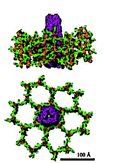
although structures are available for the homologous OM (TolC [35]) and RND (AcrB [52]) components of the Mex-like AcrAB-TolC multidrug efflux system of Escherichia coli. The TolC channel is a trimer and spans both the OM (as a ß-barrel) and periplasm (as a {alpha}-helical barrel) (35). Measuring 140 Å in length, the channel is open at the distal (extracellular) end and tapers almost to a close at the proximal (periplasmic) end, which likely interacts with the RND component, AcrB (35). Modeling studies suggest that OprM adopts a similar structure (43, 80). The AcrB RND component also exists as a trimer, composed of a 50-Å-thick transmembrane region and a 70-Å headpiece that protrudes into the periplasm (52). This headpiece has a funnel-like opening at the top that is connected to a central cavity at the bottom, which, in turn, opens to the periplasm via three vestibules that likely play a role in substrate recognition by and/or access to the pump (52). Indeed, several studies highlight the role played by the periplasmic portion of the RND transporters of E. coli and P. aeruginosa in substrate (i.e., drug) recognition (14, 15, 45, 53, 79, 83). A high-resolution crystal structure for an MFP efflux component is not yet available, although preliminary studies indicate that, e.g., AcrA is also likely trimeric (85) and that monomer AcrA is a highly asymmetric, elongated molecule of sufficient length to span the periplasm (4, 84).


Koronakis, V., A. Sharff, E. Koronakis, B. Luisi, and C. Hughes. 2000. Crystal structure of the bacterial membrane protein TolC central to multidrug efflux and protein export. Nature 405:914-919.


Bold indicates the words that provide leads to interesting aspects of secretion by Pseudomonas.

A mini-review essay topic could well focus entirely on homoserine lactone or maybe protein virulence factor secretion by Pseudomonas, and could involve careful reading, and re-reading, of 2 to 3 relevant review papers and 4 to 6 good research papers on the topic. That's enough work for an assigment.

PS Remember to ITALICISE all those species binomials in your final assignment. Cite references professionally too.

And finally, trust your luck .

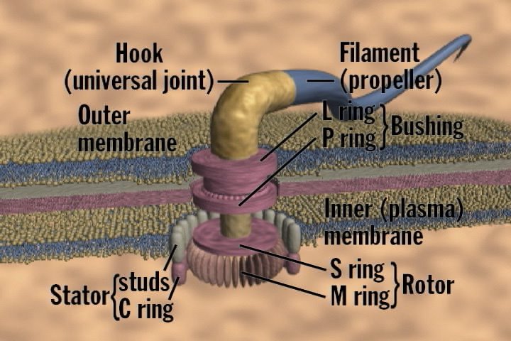
In an effort to brighten up this post with a colourful image (posted at the top), I did a google image search for bacterial multi-drug efflux pumps and found this.

Discussion thread on Cell Division in Bacillus and Caulobacter.

Discussion thread on cell division in Bacillus subtilis and Caulobacter crescentus
This is designed to help with an assignment.

Put your questions and findings in the comments please.

Cell division is one of the most rapidly developing and exciting areas of bacteriology today .

How can a novice find out about it and do literature research on the topic for a short review of what is currently known?

The first professional step is to find a high quality recent scientific mini-review in the literature.

First go the NCBI-PubMed literature search engine here.

(If in doubt google NCBI and press the PubMed button.)


Type in something like this,

cell division Caulobacter review

and press enter (or return).

A literature search output will appear on the screen. Be aware there are various options for display and textdownloading. The full summary option is most useful. Buying a cheapish USB thumb-drive to help harvest this information will be a good investment too.

You get this high up on the output list

Trends in Cell Biology
Volume 15, Issue 7 , July 2005, Pages 343-345

doi:10.1016/j.tcb.2005.05.002 Research Focus

Bacterial DNA segregation by the actin-like MreB protein

Thomas Kruse and Kenn Gerdes E-mail The Corresponding Author

Department of Biochemistry and Molecular Biology, Campusvej 55, DK-5230 Odense M, University of Southern Denmark, Denmark

Available online 25 May 2005.


Faithful chromosome segregation is vital to all organisms. Eukaryotic cells use the tubulin-based cytoskeleton to segregate their chromosomes during mitosis. A handful of papers have provided convincing evidence that, in bacteria, this task is accomplished by the actin homolog MreB. In particular, a recent study by Gitai et al. demonstrates that MreB specifically binds to and segregates the replication origin of the bacterial chromosome.


Before we go on, note that the PubMed outut for this article has a Related papers hyperlink button. Pressing it will virtually complete your assignment research for you.

Now go to a good library and search for the full copy of the journal article. An electronic version is best, as you can cut and paste your own notes onto your brand new thumdrive just as I do here.

In the mini-review full text article you find all sorts of goodies about Bacillus Caulobacter and MreB

(Note MreB has been mentioned in the lectures.)

Microbe Pundit found this :

Could MreB form a bacterial mitotic-like machine?

It is now clear that bacteria contain true homologs of both tubulin (FtsZ) and actin (MreB and ParM). Could cytoskeletal elements contribute to DNA segregation in bacteria? Indeed, the DNA segregation machinery encoded by the E. coli plasmid R1 specifies a simple prokaryotic analog of the eukaryotic spindle apparatus. The plasmid-encoded ParM protein, an actin homolog, forms F-actin-like filaments that are responsible for the active movement of plasmid copies to opposite cell poles [Ref] 5 and 6. The chromosome of rod-shaped bacteria encodes another actin homolog called MreB. The structure of monomeric MreB is very similar to yeast actin (Figure 1), thus placing the evolutionary root of actin in the prokaryotic domain [7].

[...]

In addition to its role in cell shape determination, results from several research groups have linked MreB to a function in chromosome segregation. Expression of mutant forms of MreB with impaired ATPase activity in otherwise wild-type E. coli produced rod-shaped cells with unevenly distributed and abnormal MreB filament morphologies, leaving parts of the cells with no detectable MreB signal. Under these circumstances, severe chromosome segregation defects were observed [10]. Consistently, depletion of MreB in both B. subtilis and C. crescentus leads to a rapid defect in chromosome segregation, where replication origins fail to localize in a regular bipolar fashion 17 and 18. Together with the observation that MreB forms dynamic filaments that move away from the mid-cell towards opposite cell poles in B. subtilis, these result indicate that MreB could be part of a mitotic machinery involved in chromosome segregation 4 and 13.

[...]
Inhibitor confirms role for MreB in chromosome segregation

Recent convincing evidence for a direct role of MreB in chromosome segregation in Caulobacter crescentus was presented by the Shapiro laboratory [19]. They used a small molecule, S-(3,4-dichlorobenzyl)isothiourea (A22), that was originally identified in a screen for compounds that induce anucleate E. coli cells and a change in cell morphology from the normal rod-shape to a spherical form [20]. When administered to Caulobacter cells, A22 mimics the effects of MreB depletion and causes a rapid and reversible disintegration of the MreB cables.

[...]

9 L.J. Jones et al., Control of cell shape in bacteria: helical, actin-like filaments in Bacillus subtilis, Cell 104 (2001), pp. 913–922.

11 R.M. Figge et al., MreB, the cell shape-determining bacterial actin homologue, co-ordinates cell wall morphogenesis in Caulobacter crescentus, Mol. Microbiol. 51 (2004), pp. 1321–1332.

I have bolded portions of the text which are pure gold nuggets for an assignment.
If I had time I would mention them in a lecture.

Note how two major concepts developed the classroom, MreB "cables" and chromosome movement/partition are connected to one-another by this review. This field is surely moving fast.

Note also the quality of writing and the currentness of the information in the mini-review. It is a fine example of an assigment that I would award an 110% grade (or more).

Monday, March 06, 2006

Using flow cytometry to study the bacterial cell cycle.

Diagram of flow cytometry experimental set up from here (which has extensive notes on the theory and practice of flow cytometry).


Flow cytometry is a tool for making light scattering and fluorescence emission measurements on individual cells. The equipment can collect data from numerous individual cells passing through the detector and present the collected data graphically. Usually light scattering is measured as an indicator of cell size and fluorescence staining as a measure of some component, such as stained DNA or amount of bound antigen specific antibody that is detectable by fluorescence because it has been coupled chemically to a dye.


Chapter 9 of Schaechter presents data of this type in Figure 9.3.

Fig. 9.3 describes a bacterial culture (probably E. coli) that has been treated with antibiotic rifampin, and then allowed 90 minutes to complete DNA replication before being analysed by flow cytometry.

Study Questions:

What is the target of action of rifampin?

What is the basic model for chromosome replication in these cells?

How can cells have 4 chromosomes?

How do you think DNA content per cell was measured?

Friday, March 03, 2006

Making a Cell Part 2


Adeninine related nucleotides including ATP, and NAD(P), shown here, are important coupling compounds in metabolism


One of the important roles of Fueling reactions is to generate chemical energy for cellular activities.

This energy is in the form of either ATP or PMF.

Energy sources ATP and PMF are intra convertible by cells.

This energy can be used for either Growth related activities or Growth independent activities.

Another major fueling reaction is production of reducing power (carried by NAD(P) as Hydrogen atoms attached the nicotinamide ring) which is also consumed in growth-related processes.


Study questions:

What are examples of non-growth related processes that consume energy?


Quantitatively speaking:

What are the main growth related processes that consume ATP?

What are the main growth related processes that consume reducing power?

Making a cell.

The broad processes of bacterial metabolism (fueling, biosynthesis, polymerisation, assembly, and cell division) are interrelated by the fact that each requires as a starting material the products of the preceding phase.

Study Questions:
For each phase list the starting materials and products.
(No more than one phase answered per student, to spread out the activity.)


Could any phase be dispensible? If so under what circumstances?

Operation and assembly of the bacterial flagellum.

Image from here.


Chapter three describes the three main parts of a flagellum.

Basal body, hook and filament.

The filament is hollow and made of flagellin, and rigid.The flagellin subunits assemble spontaneously to give the filament.

The hook is at the base of the filament and appears to act like a "universal joint" when the flagella is rotating.

The basal body is imbedded in the cell membranes, and include some 15 different proteins.

It acts as an electric rotating motor driven by PMF.

The flagellum is assembled in a stepwise fashion. First the basal body is inserted into the membrane, next the hook, finally the filament.

Units of flagellin reach the tip through its hollow centre.

Regulatory Loops.
In E. coli synthesis of flagellin subunits is coupled to their assembly at the tip by a special inhibitor of flagellin synthesis. When the basal body is inserted into the membrane it accomplishes secretion of this inhibitor into the growth medium which thus allows filament subunit synthesis to start.

Proteins needed to allow the flagella to rotate are added late in organelle assembly.

Study Questions:

What is PMF and where does it come from?

How were the stages of flagellum assembly first identified?

Wednesday, March 01, 2006

Protection of the cell membrane

Protection of the cell membrane in Gram positive and Gram negative bacteria is a important topic that is discussed very well in Chapter 2 of Schaechter 2006.


How do these two different types of bacterial outer cell layers (G+ versus G-) exclude toxic hydrophobic compounds?

How do hydrophilic compounds less than 700 molecular mass enter Gram negative cells?