Tuesday, October 31, 2006

A question of cycles of oxidation in mineral leaching.

Question from student (edited):

Regarding acid mine drainage, does the oxidation of sulfides to H2SO4 result in the release of the metal ions which get leached out (eg, the release of Fe2+ from FeS2)? Does the H2SO4 that is produced cause the leaching out of other metals from their ores because of its acidic nature? Can the microorganisms also oxidise the S2- in the FeS2 in addition to oxidising the Fe2+ that is produced from this reaction?

Answer (from Pundit's Environmental Expert Jan Pundit):

Initially, the H2SO4 formed by microorganisms helps to solubilise metals released from the ores. They oxidise the S2- (in the metal sulfides) to produce the H2SO4, which helps to stabilise Fe2+ (which oxidises in air under neutral or alkaline conditions), and keeps Fe3+ in solution, which would otherwise react with water to produce ferric oxy-hydroxides (rust) at neutral and alkaline pH. Once the system is acidic, and there is a high concentration of Fe3+, then the propagation cycle becomes important. There is direct chemical oxidation of the sulfides in the ores, by Fe3+, to produce more H2SO4, and releasing more (reduced) metal ions, and the Fe3+ is chemically reduced to Fe2+. The bacteria are now important for the biological reoxidation of Fe2+ to Fe3+, which is required to keep the chemical oxidation going.

Labels: , , ,

Saturday, September 02, 2006

Twisted News item for students of bacterial cells: How transport proteins work.


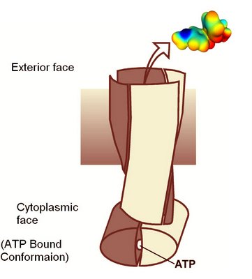
Schematic of Staphylococcus membrane transport protein in the ATP binding conformation.

A structure an ABC family transport protein that exports dyes and drugs from Staphylococcus aureus has just been reported in the journal Nature. The key discovery is that the two protein dimers in the molecule are intimately twisted around each other. The structure provides many important clues on how the transport mechanism is driven by ATP hyrdrolysis.

Structure of a bacterial multidrug ABC transporter

Roger J. P. Dawson and Kaspar P. Locher
Abstract
Multidrug transporters of the ABC family facilitate the export of diverse cytotoxic drugs across cell membranes. This is clinically relevant, as tumour cells may become resistant to agents used in chemotherapy. To understand the molecular basis of this process, we have determined the 3.0 Å crystal structure of a bacterial ABC transporter (Sav1866) from Staphylococcus aureus. The homodimeric protein consists of 12 transmembrane helices in an arrangement that is consistent with cross-linking studies and electron microscopic imaging of the human multidrug resistance protein MDR1, but critically different from that reported for the bacterial lipid flippase MsbA. The observed, outward-facing conformation reflects the ATP-bound state, with the two nucleotide-binding domains in close contact and the two transmembrane domains forming a central cavity—presumably the drug translocation pathway—that is shielded from the inner leaflet of the lipid bilayer and from the cytoplasm, but exposed to the outer leaflet and the extracellular space.
Nature advance online publication 30 August 2006 | doi:10.1038/nature05155; Received 9 May 2006; Accepted 11 August 2006; Published online 30 August 2006

Labels: , ,

Tuesday, August 29, 2006

Entry of dyes can be sensitive to the state of the membrane

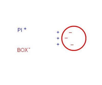
Schematic diagram of a living (bacterial) cell with its polarised (charged) membrane. The cytoplamic membrane is represented in red. PI is a cationic dye (propidium iodide) that binds to DNA, and it is not lipid soluble. BOX is lipophilic oxanol dye, negatively ionised, and about 500 MW. Both PI and BOX are fluorescent.


Question: What are the factors affecting staining of the cell by these dyes?

Labels: , ,

Monday, May 22, 2006

Why do Streptomycetes have ability to produce so many specialised molecules?

Various streptomycete soil bacteria have ability to produce a range of molecules called secondary metabolites.

See if you can relate a specialised molecule, or streptomycete general metabolic versatility, to one of the following aspects of streptomycete biology.

  1. Habitat
  2. Life-cycle
  3. Cell fusion between different vegetative hyphae
  4. Presence of conjugative plasmids
  5. Nutrition and nutrient capture
  6. Chromosomal structure
  7. QS
  8. Membrane physiology
  9. Stress - UV exposure, dehydration
  10. Aerial hyphae
  11. Spore survival

Good luck.

Labels: , , ,

Wednesday, February 22, 2006

Microbe. The World of Microbes.


As a start to this discussion forum, it's good to go to the website devoted to book Microbe that inspired this weblog. This website is here.

We are starting discussions with chapter 1 of this book- The World of Microbes.

Let's get started by posting a study question for discussion in the comments thread.

Question
Bacteria range in volume over a million-fold. Discuss some of the consequences of being larger or smaller than average?

Update of question and posting with more information to assist students. 4th March 2007
Here is some extra information that can assist students understand how size has implications for available membrane surface area to service the metabolic requirement of a unit cell volume.

To understand the point of the question about size, students need to think about have the ration of surface are:cell volume changes with increase in size. SA/Vol Ratio~diameter squared/diameter cubed~inversely proportional to diameter, with similar cell shapes.

To see some consequencences raised by the biology of large bacteria you need to read about observations that have been made on Epulopiscium bacteria, usually called epulos. The paper by K D Clements and S Bullivant (1991) An unusual symbiont from the gut of surgeonfishes may be the largest known prokaryote. J Bacteriol. 1991 September; 173(17): 5359–5362 provides some good interesting insights.

Figure 1 of this paper shows how large epulos are.
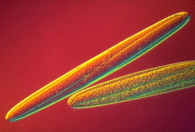
J Bacteriol. 1991 September; 173(17): 5359–5362. Figure 1. Light micrograph of an Epulo. The letter C indicates a smaller, eukaryotic ciliate.

J Bacteriol. 1991 September; 173(17): 5359–5362. Figure 3. Electron micrograph of a thin section of an Epulopiscium bacterium showing concolutions to cytoplasmic membrane. Such convolutions would increase the membrane area to cell volume ratio of these large bacteria

Thus epulos have a peripheral layer of highly convoluted cytoplasmic membrane - which has been interpreted as a mechanism increase in membrane area to compensate for some surface area to volume related challenges that they face because of their large size.

Extra reading for the high achieving student:
In the links at the Microbe Chapter 1 webpage, this is worth studying several times in the course.

Labels: , , ,