Tuesday, October 31, 2006

Q and A on BACs and Metagenomics. Why large inserts?

Question from student (edited):

I am reviewing the Soil Metagenome paper that we reviewed in class (Rondon et al., 2000, Appl. Environ. Microbiol. 66: 2541-2547), and have a written note claiming that there are two important reasons to clone large DNA fragments into the BAC vectors. I can only think of one reason, which is that many soil microbes produce important bioactive compounds, and genes required for production along with regulatory genes are often clustered in one continuous segment on the chromosome called an operon. A large fragment of DNA inserted into BAC plasmid would increase the likelihood that the entire operon included. Is this correct, and is there another reason why having large DNA fragments in the BAC plasmids is important?

Answer:

Yes, you are correct, and yes, there is another reason. Large inserts mean that each clone (Escherichia coli clone line) is likely to contain genes encoding for multiple activities that may be of interest. This means that fewer clones need to be maintained than if the inserts were smaller, i.e., the same amount of metagenomic information is maintained in fewer clones (or more metagenomic information can be maintain in any given number of clone lines). Using screening assays for different activities, the same amount of metagenomic material can therefore be interrogated for the presence of specific activities while manipulating fewer cultures.

Labels: , , , ,

A question of cycles of oxidation in mineral leaching.

Question from student (edited):

Regarding acid mine drainage, does the oxidation of sulfides to H2SO4 result in the release of the metal ions which get leached out (eg, the release of Fe2+ from FeS2)? Does the H2SO4 that is produced cause the leaching out of other metals from their ores because of its acidic nature? Can the microorganisms also oxidise the S2- in the FeS2 in addition to oxidising the Fe2+ that is produced from this reaction?

Answer (from Pundit's Environmental Expert Jan Pundit):

Initially, the H2SO4 formed by microorganisms helps to solubilise metals released from the ores. They oxidise the S2- (in the metal sulfides) to produce the H2SO4, which helps to stabilise Fe2+ (which oxidises in air under neutral or alkaline conditions), and keeps Fe3+ in solution, which would otherwise react with water to produce ferric oxy-hydroxides (rust) at neutral and alkaline pH. Once the system is acidic, and there is a high concentration of Fe3+, then the propagation cycle becomes important. There is direct chemical oxidation of the sulfides in the ores, by Fe3+, to produce more H2SO4, and releasing more (reduced) metal ions, and the Fe3+ is chemically reduced to Fe2+. The bacteria are now important for the biological reoxidation of Fe2+ to Fe3+, which is required to keep the chemical oxidation going.

Labels: , , ,

Analogue resistant mutants and the discovery of metabolic regulation are inextricably linked in history.

The History of Using Metabolic Analogs to Reveal Regulation of Metabolism.


Metabolic analogues
are compounds that structurally resemble natural metabolites.

A name for a metabolic analogue that interferes with cell functions is antimetabolite.

Antimetabolities will often inhibit cell growth.

Sulfa drugs were perhaps the first antimetabolites.


In 1958, Ed Adelberg discovered that Escherichia coli MUTANTS, that had been selected as being resistant to growth inhibition by toxic metabolic analogues, secreted excess quantities of certain metabolites into their growth medium that the wild-type parental strains DID NOT secrete.


For instance, E. coli mutants selected for resistance to p-fluorophenylalanine secreted tyrosine and only tyrosine into the medium.

p-Fluorophenylalanine is a tyrosine analogue.

The compounds secreted by E. coli mutants differed when different types of antimetabolite were used to select mutants.

Ethioninione resistant mutants secreted methionine. (The structure of ethionine is Ethyl replacing the Methyl in methioninine.)

The patterns of secretion suggested to Adelberg that resistance was based on COMPETITION between metabolite and analogue.

Interestingly, metabolic analogues were provided to Ed Edelberg by Arthur Pardee, one of the discovers of feed-back inhibitable biosynthetic enzymes (allosteric enzymes).

In 1960 Pardee reported 3-Methylaspartic acid as a potent antimetabolite of aspartic acid in pyrimidine biosynthesis, showing these ideas are not confined to amino acid pathways.

Selection of bacterial mutants which excrete antagonists of antimetabolites.
J Bacteriol. 1958 Sep;76(3):326.Click here to read Links
ADELBERG EA
Now marvellously available online via Pubmed
http://www.pubmedcentral.nih.gov/articlerender.fcgi?tool=pubmed&pubmedid=13575393

Famously, Pardee and Yates had discovered feedback-inhibition in 1956, in a key paper revealing the existence of biochemical regulation.

Dick Yates went on to work in applied microbiology in Wilmington DE, and the Pundit is honoured to have met this gracious American scientist.

J Biol Chem. 1956 Aug;221(2):757-70.
Control of pyrimidine biosynthesis in Escherichia coli by a feed-back mechanism.
PARDEE AB, YATES RA.
PMID: 13357469 [PubMed - OLDMEDLINE for Pre1966]
http://www.jbc.org/cgi/reprint/221/2/757

Related Links
Pyrimidine biosynthesis in Escherichia coli. [J Biol Chem. 1956]
PMID:13357468
Studies on the biosynthesis of bacterial and viral pyrimidines. IV.
Utilization of pyrimidine bases and nucleosides by bacterial mutants. [J Biol
Chem. 1957] PMID:13475346
3-Methylaspartic acid as a potent antimetabolite of aspartic acid in
pyrimidine biosynthesis. [J Biol Chem. 1960] PMID:13786646

Labels: , ,

Monday, October 30, 2006

Discovering novel antifungal agents with antimitotic activity from diverse soil and water sources.

C W S Fen, Melbourne

Abstract
In response to the side effects and increasing amount of resistance met by antifungal drugs targeting the ergosterol pathway in fungi, we propose to bring antimitotic antifungal agents to a reformation, enlisting the use of high throughput screening to discover novel microtubule-inhibiting compounds produced by fungi from a diverse range of soil and water sources. We intend to screen large amounts of secondary metabolites produced by these fungi against 10 specifically chosen common infection-causing fungi species. After narrowing down the candidate compounds to a few lead compounds, these will be tested for fungal-specific antimitotic activity in vitro with fungal microtubule dimers and in vivo against the 10 fungi species utilizing GFP-labelling technology. Toxicity to human cells will be assessed as well before clinical trials can be proposed. Further experiments are necessary to determine optimum dosage and method of delivery of the drug, and further research into potential anticancer drug applications may yield additional commercial benefit from investment in this initiative.

Introduction
Imagine if a harmless scab that you thought would just go away became the cause of your hospitalisation. Or the joy and relief of being discharged after an operation only to find yourself back in bed being treated for a post-surgical fungal infection. Fungal infections have a significant impact on the quality of life of affected individuals despite little attention dedicated to it when compared to other life-threatening diseases. However, most predominantly, it is patients who have undergone surgery, are immunocompromised, have undergone chemotherapy or excessive antibiotic doses who are severely affected and whose life may well be in danger if the infection is not addressed immediately (White et al., 1998). In view of this, biotechnological innovations and advances over the past few years can be put to good use in fuelling the initiative of discovering new or improved anti-fungal compounds which targets fungi specifically while sparing human cells.

In light of this, we propose to bring fungal-specific antimitotic drugs to a reformation, achievable by exploiting the fungal cells’ dependence on microtubule function to proliferate, and thus prevent the progression of infection. From a marketability standpoint, the diverse range of pharmaceuticals that target the ergosterol pathway in fungi are saturating the market, resulting in resistance among different drugs targeting this same biochemical pathway (White et al., 1998). Therefore the guaranteed effectiveness of this new drug minus the side effects of its competitors (which are the main source of concern), coupled with increased efficacy, is highly promising in guaranteeing a high return in investment. Accordingly, we intend to perform high-throughput screening of soil and water samples from a diverse range of sources to screen for fungi that have the ability to produce such a compound. Following that, we will run carefully designed assays to engineer additional broad-spectrum activity of the said compound. Miracles are more likely to happen if we make them and we intend on doing just that.

PROPOSAL:

The Scientific Basis

Microtubules are essential to all eukaryotic cells; in essence, fungal cell shape, transport, motility and most importantly for this proposal, cell division, rely wholly on the proper function of this critical organelle. The fundamental property of microtubules that we look to exploit in a biotechnological sense is its non-equilibrium behaviour, better known as microtubule dynamic instability, which determines the shortening or lengthening of microtubules to facilitate them in performing the functions listed (Nogales 2001). With this in mind, we analysed fungal cell characteristics to determine a logical strategy of attack.

Many secondary metabolites produced by fungi have potent pharmacological uses through exploiting its initial intended function. An obvious example of this is Penicillin produced by the mould Penicillium notatum a commonly used antibiotic for treating bacterial infections and diseases. However, this same species of mould from which penicillin is derived also produces another compound, Griseofulvin, synthesised by a close relative, Penicillium griseofulvum, which prevents microtubule formation in fungal cells during mitosis. Although griseofulvin requires a longer duration of action and exhibits less efficacy than other commonly used antifungals, nevertheless, it boasts minimal side effects and a currently still unexplored biochemical mechanism as to why it targets fungal cells specifically, partially sparing human cells and therefore resulting in milder side effects (Jimenez et al, 1990). Logically assuming that it is the specific action or higher affinity of the drug for fungi microtubules that results in the milder side effects, we propose to further expand on the merit of this particular target protein of the drug and search for novel compounds which utilises a similar mechanism of attack to halt the growth of fungi, making it a potent fungistatic agent. Subsequently (not covered by this proposal), chemical modification or further subjection of the fungi species of interest to selective chemicals or mutagenic agents can be run to develop a compound that will bind irreversibly to the microtubules and thus effectively kill off the target fungal cells (Refer to Figure 1 and Figure 2 below).

Figure 1: Two possible models of microtubule nucleation. (a) End-on model of nucleation; (b) Lateral interaction of γ-Tubulin protofilament with αβ-Tubulin dimers. (Modified from Nogales, 2001). Irreversible binding of the antifungal compound (in red – whether to the protofilament as whole or to individual dimers) can prevent prolongation of αβ-Tubulin protofilament or interaction between filaments, disrupting microtubule instability and therefore mitosis.


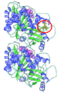
Figure 2: Ribbon diagram of molecular structure of α- and β-tubulin dimer of microtubules. A compound that can bind irreversibly to a point on this most basic unit of microtubule structure at e.g. the taxol binding site (circled in red) can trap these tubulin dimers and cause stabilization of microtubules, preventing mitosis (Revised from Nogales, 2001).


The Source

We plan to run high throughput screening of a range of soil and water samples obtained from diverse geographical locations around the world to isolate as many species of fungal cells as possible, with a more bias focus on mycoparasites, as toxic compounds produced by fungi against other fungi will be more specific and therefore more effective. Then, we will narrow down the candidates of these fungal species to those that may harbour antimitotic activities.

By diluting the soil sample in water and partially diluting the water samples, multiple cultures (100 odd petri dishes) of fungi are obtained using growth media that will stimulate fungal growth, e.g. YES media (Torres et al., 1987). After this primary culture, each fungal colony will be extracted and cultured at diluted concentrations in zone-based bioassays overlaid with a layer of one of 10 designated common infection-causing fungi, i.e. the Tinea spp., Candida spp., Aspergillus sp., Cryptococcus spp., Fusarium spp., etc. (Mays et al, 2006). It was shown by Panda et al., 2005, after a few weeks, any fungal colony whose surrounding cells are trapped in interphase and highly mononuclear exhibits antimitotic activity. These colonies are isolated and the compound produced by each colony is extracted and purified by chemical means. These compounds are then subjected to high throughput screening to test for antimitotic activity.

High Throughput Screening

The screen we have in mind is designed to be a fungal-specific antimitotic screen. Exploiting the success of genomic sequence technology and the completion of function determination of thousands of genes, we will search for genes encoding microtubule function in the 10 species of common infection-causing fungi using the available genome libraries online. By ensuring that the 10 chosen species of fungi have been studied intensively enough to for us to genetically manipulate the microtubule and its associated proteins’ genes, hypersensitive mutants susceptible to antimitotic drugs will then be generated and these mutant fungi will be used to screen for compounds that can inhibit their growth among the potential candidate compounds produced by the initial fungal culture. Testing will be done at increasing concentrations of antimitotic/antifungal compounds. Following positive results, the wild type strain for each of the 10 common disease causing fungi is then exposed to these 10 compounds as a control to identify any of the compounds can arrest the cell cycle of the parent strain (Lila et al, 2003). In this way, the antimitotic drug-sensitive fungi will increase the likelihood of discovering a drug that has antimitotic potential which we can improve the properties of through further screening.

Thus, narrowing down the search to one lead compound, we will proceed to test this in vitro with microtubules obtained from fungal cells. By tagging the fungal tubulin dimer proteins with GFP tags, we will then monitor the action of the compound on the polymerization of individual tubulin entities with increasing lead compound concentration.

From in vitro studies, we will extrapolate success of the compound in inhibiting microtubule function to in vivo testing by a reporter system where cloning technology is used to introduce fluorescence into microtubule dimers by incorporating the GFP gene into the tubulin gene (cf. protein) of each of the 10 fungal species which is then transcribed into tubulin dimers that will fluoresce, thus enabling us to monitor the growth or shrinking of microtubules (mitotic spindle) during mitosis (Implication of concept by Kumagai et al., 2003 and Alberts et al., 2002). Further proof can be obtained by running flow cytometry analysis of fungal cells treated with cytotoxic concentrations of the lead compound – if there is no increase in nuclear DNA content, then there is a lack of tubulin-related activity in vitro (Lila et al, 2003).

These same set of in vitro and in vivo experiments are repeated with human tubulin and in human cells from various tissues across the body respectively to test for toxicity of the compound (Priestly and Brown, 1978). Thus, if luck it working on our side, we may be able to determine a distinct fungal specificity of the drug and determine the exact concentration of the compound which is the threshold of toxicity for human cells. Complicated but necessary further experimentation should be done in whole organism model systems such as rats, mice and other mammals to mimic possible side effects in humans before proposing the extrapolation of these results to humans in clinical trials.

Potential Problems

Microtubules, despite being in different organisms, are highly conserved organelles, especially between organisms of the same domain (Eukaryota). We are taking a significant risk in gambling with nature on the possibility that antifungal compounds produced by fungi themselves against their fungal counterparts might specifically inhibit these competitors for survival antimitotically. At the very least however, we can design these novel compounds specifically for a distinct group of fungal species (e.g. specifically one of superficial, subcutaneous, systemic or opportunistic infection-causing groups of fungi) if not for broad-spectrum activity against most of them.

As in any microbial biotechnology process, transition from a small-scale experiment to culturing of these fungi of interest in the large scale will present us with many more problems to come. However, given the appropriate equipment, competent technicians and proficient biotechnologists, this should be a challenge we can overcome.

Further Research

As part and parcel of an antifungal treatment, side effects are inevitable as human cells are very similar structurally to fungal cells, as mentioned earlier. Thus, drugs that target fungal cells will affect our cells to a certain extent as well. Therefore, although the side effects will be specifically tailored to be minimal, further chemical modification or manipulation (if possible) to obtain compounds that have near-negligible side effects will be needed following the isolation of the fungal microtubule inhibitor protein-producing fungi..

In addition to that, also on a pharmacological basis, we need to take into account the stability of the compounds (determining optimal pH), scaling up of production and the efficiency of cell growth of the cell of interest in culture. Other than that, solubility of antifungal compounds is a major concern in the pharmaceutical industry as most antifungal compounds are contrary to that requirement. Added to that oral bioavailability, potential side effects and possible allergic reactions have to be accounted for to guarantee its economic value.

Potential Future Benefits (Integration of Science and Business)

One of the key valuable gains from the production of this initial microtubule formation inhibitor compound is a better understanding of the mechanism of microtubule inhibition and prevention of mitosis. From investigating so many fungi species with antimitotic activity, we can gain an insight into the many potential ways the microtubule can be inhibited and by manipulating this knowledge as well as other biotechnological screening and assay methods to slightly modify the compound, we could extrapolate the research to involve discovery of similar compounds from fungi which will specifically inhibit microtubule formation of tumour cells and thus secure a spin-off opportunity to produce a potential anti-cancer drug. Much research efforts and funds have been poured into a similar initiative involving Griseofulvin (Panda et al., 2005). Thus, this presents a further bonus for investing in this research.

Conclusion

In conclusion, this initiative of discovering novel antimitotic antifungal drugs presents many commercial, scientific and medical benefits to every player: investors, consumers and the biotechnologists. Besides solving a demanding medical problem and gaining much knowledge about fungi and its infection-causing mechanisms, there is much to gain from investment in the long run, with spin-off opportunities that are becoming a reality in the anticancer drug industry. “Fortune favours the prepared mind - Louis Pasteur.” This statement could not hold truer in this fascinating hybrid between business and science.

References
1. Lila, T., Renau, T.E., Wilson, L., Philips, J., Natsoulis, G., Cope, M.J., Watkins, W.J. and Buysse, J. 2003. Molecular Basis for Fungal Selectivity of Novel Antimitotic Compounds. Antimicrobial Agents and Chemotherapy. 47: 2273-2282.

2. Nogales, E. 2001. Structural Insights into Microtubule Function. Annual Review of Biophysics and Biomolecular Structure. 30:397-420.

3. Kumagai, F., Nagata, T., Yahara, N., Moriyama, Y., Horio, T., Naoi, K., Hashimoto, T., Murata, T. and Hasezawa, S. 2003. Gamma-tubulin distribution during cortical microtubule reorganization at the M/G1 interface in tobacco BY-2 cells. European Journal of Cell Biology. 82:43-51.

4. Alberts, B., Johnson, A., Lewis, J., Raff, M., Roberts, K. and Walter, P. 2002. Molecular Biology of The Cell, 4th edition. Garland Science, New York, USA.

5. White, T.C., Marr, K.A. and Bowden, R.A. 1998. Clinical, Cellular, and Molecular Factors That Contribute to Antifungal Drug Resistance. Clinical Microbiology Review. 11: 382-402.

6. Torres, M., Canela, R., Riba, M. and Sanchis, V. 1987. Production of patulin and griseofulvin by a strain of Penicillium griseofulvum in three different media. Mycopathologia. 99: 85-89.

7. Mays, S.R., Bogle, M.A. and Bodey, G.P. 2006. Cutaneous Fungal Infections in the Oncology Patient: Recognition and Management. American Journal of Clinical Dermatology. 7: 31-43.

8. Priestly, G.C. and Brown, J.C. 1978. Effects of griseofulvin on the morphology, growth and metabolism of fibroblasts in culture. British Journal of Dermatology. 99: 245.

Labels: ,

Step up: Bacterial Lipase to Substitute Pancreatic Lipase For Enzyme Therapy

D. G. Sani, Melbourne

Summary
Microbial lipases have been more commonly used than lipases derived from plants and animals due to higher stability, rapid production, higher yields and more convenient manipulations of microorganisms. For example, lipase from Mucor miehei is used in cheese production, Candida antartica in surfactants, Serratia marcescens lipase in drugs (diltiazem). Medical area is another promising area where lipase can be used as digestive aids. To date, only pancreatic enzyme (lipase) therapy is used to treat fat malabsorption in Cystic Fibrosis and pancreatitis patients. However, pancreatic lipase is susceptible to low pH (acidic gastric environment) and protease which can render the lipase inactive. Therefore, it is proposed that an alternative bacterial lipase might have the ability to retain its activity under acidic condition and also be protease tolerant. Screenings for this novel bacterial lipase would be done from bacterial populations that are known to survive in the gastric environment. Moreover, different assays would be done to observe the lipase production, activity, and specificity under certain conditions.


A. INTRODUCTION

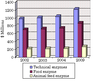
Bacterial lipases have been recognized since nearly 100 years ago in lipase-producing bacteria such as Bacillus pyocyaneus (today is named Pseudomonas aeruginosa), Staphylococcus pyogenes (S. aureus), B. fluorescens (P. fluorescens) [1]. For a lipase to be defined as a true lipase, it must exhibit interfacial activation where a presence of triglyceride should rapidly increase its activity; it should also contain a loop allowing substrate entry to the active site. However, these two criteria are unsuitable for some exceptions where the lipases have the loop but do not exhibit interfacial activation. Hence, a simpler definition of lipases is carboxylesterases that catalyse the hydrolysis and synthesis of long-chain acylglycerols [1]. The prospect for industrial enzymes (technical, food, and animal feed enzyme) is very promising with an estimated increase to $2.4 billion in 2009. Technical enzyme such as in detergents, pulp production, and medical treatment is predicted to have the highest share at 52% [2].


Global Enzyme Markets Based on Application Sectors, 2002-2009
($ Millions)

Source: BCC research [2]


Application of lipase in industrial enzyme production has covered a wide area: flavour development in food technology; as oil removal in detergents; textile making; personal care products e.g. cosmetics; digestive aids for medical treatment [3]. As digestive aids, pancreatic lipase has been widely used in the enzyme therapy to treat lipid malabsorption e.g. in cystic fibrosis and pancreatitis patients. However, there are some barriers with the pancreatic enzyme therapy that prevent its maximal efficacy [3]. Hence, the need of finding an alternative lipase from microorganisms has been raised. Microbial enzymes have several advantages than animals or plants enzymes due to the stability, high yields and rapid production from microbes, and variety in catalytic activity [3]. This paper will firstly describe the background and target market of enzyme therapy for lipase, followed by proposal of methods to isolate lipase-producing bacteria and assays to obtain the acid and protease tolerant lipase.


B. BACKGROUND AND TARGET MARKET
Cystic Fibrosis (CF) is an autosomal genetic disorder affecting mostly Caucasian populations with approximately one in every 3500 newborns is born with CF each year in the United States alone [4]. Being a multi-system disease, CF affects many organs: lungs, pancreas, liver, bones (osteoporosis) [5]. The altered gene in CF disrupts the function of salt transports in organs leading to an excess production of thick, sticky mucus that blocks the ducts in these organs mainly the lungs and the pancreas. Patients with affected pancreas tend to have digestion and growth problems due to pancreatic insufficiency where there is a lack of pancreatic enzyme produced by the body [4,5]. This condition leads to lipid malabsorption because the body cannot break down the essential fatty acid (EFA) molecules coming from external nutrients [6]. Poor nutritional status due to EFA deficiency is correlated with weight and height retardation in children with CF. Moreover, a research by CDC in the US has shown a correlation between nutritional status and the lung function showing increase in body weight in CF patients (measured by BMI) is accompanied by increase in the lung function [4].

Many treatments have been developed to treat the malnutrition in CF patients and improve their life expectancy. To date, pancreatic enzyme (lipase) therapy from porcine origin has been the most widely used since 1930s with about 90% of the patients taking pancreatic enzyme supplements in 2004 [4, 7]. However, the efficiency of this treatment has not reached maximal yet. The pancreatic lipase is found to be denatured by gastric acids before reaching the small intestine where the FA hydrolysis and absorption mainly occur. Although coating the enzyme with an acid resistant (enteric) coating appears to improve the delivery of the enzyme into the small intestine, the low intestinal pH in CF patients after meal (pH less than 4) would still irreversibly denature the pancreatic lipase [7].

Another barrier for the existing pancreatic enzyme therapy is that pancreatic lipase is also denatured by proteases especially chymotrypsin in the intestinal lumen [7]. Hence, in search of ways to improve the enzyme therapy, the aim is to find an alternative lipase that would be acid resistant, protease resistant and would still possess the pancreatic lipase activity.


C. SCIENTIFIC BACKGROUND FOR BACTERIA ISOLATION
In order to find a lipase which shares features with pancreatic lipase, one must know the characteristics of pancreatic lipase. It is known that pancreatic lipase hydrolyses long-chain triacylglyceride (TG) into FAs to be readily absorbed in the small intestine at the optimal pH of 8 [6]. When the pH falls below six the pancreatic lipase is rendered inactive and if the pH less than 4.5, it is irreversibly inactivated; the later is the case in CF patients causing denaturation of the pancreatic lipase in the small intestine during the enzyme treatment [6, 7]. Hence, the alternative lipase would have to be able to survive under acidic environment (pH less than 4). Moreover, knowledge of the active site structures of pancreatic lipase can be a valuable information in finding a new lipase. The active site residues of pancreatic lipase are serine (Ser 152), aspartate (Asp 176), and histidine (His 263) [8]. These are found to be conserved in bacterial lipases where the active site has a nucleophilic residue (Ser, Cys or Asp), a catalytic triad residue (Asp or Glu), and a histidine [1].

Acid resistant lipase could most probably be obtained from bacteria living in acidic environment. A study shows that Salmonella, a gastrointestinal pathogen, produces proteins: RpoS, PhoP, and Fur that help them survive under the harsh gastric environment. RpoS and Fur are responsible for survival against organic (weak) acid stress, while PhoP and RpoS act against inorganic acid (low pH) stress [9]. Therefore, bacteria that produce RpoS, PhoP and Fur would presumably be able to survive under low pH in the gastric environment. A database search from the genbank of NCBI returns 33 bacterial strains that have RpoS, PhoP, and Fur proteins in their genome. Most of these strains are from Salmonella, Pseudomonas, E. coli, Yersinia, Shewanella, and Shigella [10].

Out of these bacterial populations, previous studies have shown that some Pseudomonas strains produce true lipase while both E. coli and Salmonella typhimurium produce esterases [1]. Bacteria surviving from the gastric environment would most probably be present in faeces. Hence, novel lipase-producing bacteria can be screened from faecal specimens and looking specifically for Yersinia, and Shigella as no lipases have been identified from these genera before. These specific strains can be isolated from faecal specimens using selective media. For example, MacConkey agar (MAC) is used to isolate lactose-fermenting, gram negative enteric bacteria while xylose lysine desoxycholate (XLD) agar is specific for isolation of Salmonella and Shigella [11]. Yersinia can be isolated on DYS medium containing peptone, ox-bile, arginine, lysine, arabinose, casein where Yersinia will ferment arabinose and appear as bright red colonies [12].

Another consideration in searching for a new lipase is the specificity of the lipase. Since the aim is to screen for a bacterial lipase that mimics pancreatic lipase, the bacterial lipase should be able to hydrolyse long-chain TGs- the substrate for pancreatic lipase [6]. Although the term ‘long-chain’ TG is not strictly defined, in general, glycerolesters with an acyl chain length more than ten carbon atoms can be considered as lipase substrates e.g. trioleoylglycerol. On the other hand, esterase substrates would normally have an acyl chain length less than 10 carbon atoms e.g. tributyrylglycerol. Most lipases, however, are capable of hydrolysing esterase substrates in addition of the lipase substrates [1]. Hence, screening for the alternative lipase would differentiate between lipase and esterase specificities.

D. ASSAYS
Assays that would be used for screening of an alternative bacterial lipase are assays for lipase production, lipase purification, activity and specificity, pH effects on activity (low pH), and tolerance on protease (chymotrypsin).

Identification of lipase-producing bacteria from the formerly isolated strains can be done by a plate assay where the agar medium contains triolein as the lipase substrate. Lipase production on triolein plates is indicated by orange-red fluorescence of the colonies at 350 nm UV lamp [1]. The lipase-producing colonies can then be purified by inoculating a single colony onto agar plates containing essential compounds for growth. Then the strains would be grown in minimal media containing some essential nutrients for growth and enhance extracellular lipase production. It has been studied that exolipase production in Serratia marcescens can be enhanced by addition of certain polysaccharides as shown in the table below [13].

Table from Winkler, U., Stuckmann, M. (1979). Glycogen, hyaluronate, and some other polysaccharides greatly enhance the formation of exolipase by Serratia
marcescens. J Bacteriol 138: 663-670. Not displayed.

Table Source: Winkler [13].

It can be seen that laminarin exhibits the highest exolipase inducing ability followed by glycogen and pectin B, then hyaluronate. Moreover, the exoprotease production should be kept to minimum so that it would not degrade other essential proteins. Amongst these exolipase enhancers, the most suitable polysaccharide would be pectin B since it has high inducing ability with less exoprotease activity produced compared to laminarin and glycogen. Furthermore, due to interfacial activation of most lipases, addition of long-chain triglyceride such as olive oil might enhance the activity. Hence, pectin B and olive oil can be included in the medium as exolipase enhancers.

There are different assays to test lipase activity in the supernatants namely: the photometric assay and titration method [1]. The photometric assay is done using p-nitrophenylpalmitate (pNPP) as the substrate which is mixed with the supernatant [13]. Lipase activity is then monitored by detecting the hydrolysis of various p-nitrophenolesters of FAs (greater than 10 carbon atoms) into p-nitrophenol at 410 nm. Different p-nitrophenolesters would be separately included in the reaction mixture to test for lipase specificity [14]. The titration method measures lipase activity by recording the amount of NaOH used to maintain pH 8 as the fatty acid is liberated after addition of lipase solution into the supernatants [15].

Assay to screen for acid tolerant lipase can be performed by setting up a reaction mixture mimicking the gastric environment where the pH can be as low as 2.5 to 4. The synthetic gastric fluid has been widely used by mixing pepsin, lysozyme, and bile. Although the synthetic gastric fluid is more convenient to prepare, it has been demonstrated that certain bacteria are more sensitive in an ex vivo porcine gastric fluid than in the synthetic gastric fluid at a same pH of 2.5. This suggests that there are some components which are present only in the porcine stomach and can interfere with bacterial survival [9]. Hence, a more reliable assay for acid tolerant lipase can be done using porcine gastric fluid collected from live pigs instead of the synthetic gastric fluid. The pH of porcine gastric fluid can be adjusted from 3.47 to 4.15 by mixing supernatants of several stomach contents with pH ranging from 1.42 to 4.44 [9]. This mixture (synthetic or porcine gastric fluid) can then be added into the bacterial supernatants and then assayed for lipase activity (photometric assay). The acid resistant lipase would retain its ability to hydrolyse p-nitrophenylesters after treatment with the gastric fluid.

Protease tolerant lipase can be assayed by adding a specific protease into the bacterial supernatants prior to the lipase activity assay. For this experiment, chymotrypsin would be used since it is found to denature pancreatic lipase in the small intestine [7]. If successful, the bacterial lipase that retains its activity would be the protease tolerant lipase. After confirming the lipase which possesses the specific characteristics, purification of the lipase can be done by chromatographic methods where the lipase is bound to beads in a column and then eluted with corresponding buffer. For instance, extracellular lipase of Pseudomonas was purified by anion-exchange chromatography and HIC [15]. The main advantage is that purified enzymes have high activity and can be re-used in other reactions.

CONCLUSION
Technical enzymes application in various biotechnology areas has shared the highest part in the global enzymes market since 2002 [2]. One of the major areas is in medical application where pancreatic lipase therapy is used as digestive aids mostly in CF patients. However, the efficacy has not reached maximal due to degradation of the lipase under acidic condition in the stomach and small intestine of these patients [7]. Hence, searching an alternative acid-tolerant lipase from bacterial origin to substitute pancreatic lipase as digestive aids would have high market returns in the future. If successful, the novel bacterial lipase would be acid and protease tolerant where it can retain its activity at pH less than 4. Moreover, purification would increase its activity implying for higher end products yields from fewer lipase mass and hence, effective production cost.

REFERENCES

1. Jaeger, K. E., Dijkstra, B. W., Reetz, M. T. (1999). Bacterial biocatalysts: molecular biology, three-dimensional structures, and biotechnological application of lipases. Annu Rev Microbiol 53: 315-51.

2. BCC report on global enzyme market. http://www.bccresearch.com/bio/BIO030D.asp. Retrieved 5/10/2006.

3. Hasan, F., et. al. (2006). Industrial applications of microbial lipases. http://www.aseanbiotechnology.info/Abstract/21019024.pdf

4. Cystic Fibrosis Foundation. (2004). Patient registry annual data report 2004.

5. Gotz, M. H. (2002). Cystic Fibrosis literature review annual report 2002.

6. Peretti, N., et. al. (2005). Mechanisms of lipid malabsorption in Cystic Fibrosis: the impact of essential fatty acids deficiency. Nutrition & Metabolism 2:11.

7. Durie, P., et. al. (1998). Uses and abuses of enzyme therapy in cystic fibrosis. J R Soc Med 91: 2-13.

8. Olivecrona, G. et al. (1994). “Medical aspects of triglyceride lipases.” In P. Woolley & S.B Petersen (Eds.), Lipases: their structure, biochemistry and application pp.316-318.

9. Bearson, S. M. D., et. al. (2006). Identification of Salmonella enterica serovar Typhimurium genes important for survival in the swine gastric environment. Applied and Environmental Microbiology 72: 2829-36.

10. NCBI web site: http://www.ncbi.nlm.nih.gov/entrez/query.fcgi?db=Genome&cmd=search&term=RpoS,+Fur,+PhoP+containing+bacteria Retrieved 29/9/2006.

11. CDC: Ch4: Identification and isolation of Shigella. http://www.cdc.gov/ncidod/dbmd/diseaseinfo/cholera/ch4.pdf#search='isolation%20of%20shigella'. Retrieved 4/10/2006.

12. Agbonlahor, D. E., et. al. (1982). Differential and selective medium for isolation of Yersinia enterocolitica from stools. J Clin Microbio 15: 599-602.

13. Winkler, U., Stuckmann, M. (1979). Glycogen, hyaluronate, and some other polysaccharides greatly enhance the formation of exolipase by Serratia
marcescens. J Bacteriol 138: 663-670.

14. Kanwar, S. S., et. al. (2006). Purification and properties of a novel extracellular thermotolerant metallolipase of Bacillus coagulans MTCC-6375 isolate. Protein Expression & Purification 46: 421- 428.

15. Kordel, M., et. al. (1991). Extracellular lipase of Pseudomonas sp. strain ATCC 21808: Purification, characterization, crystallization, and preliminary X-Ray diffraction data. J Bacteriol 173: 4836-41.

Labels: , ,

Thursday, October 12, 2006

Another Question for the crowd to kick around.

Update 30th October
Thanks for putting in questions from the list given in the study Question list not posted here.

THIS IS THE PLACE WHERE I ANSWER THEM, and will happily answer any more that are placed in the comments.

-------

Compare and contrast fructose syrup and steroid manufacturing processes as used in industry.


Both compare and contrast please.

Labels: ,

Questions 2 and 3 for discussion.

Explain why corticosteroids are relatively cheap drugs, and why fructose syrup is a cheap sweetener.

Labels: ,

Question for discussion 1

Explain the differences in use of alpha-amylase and glucoamylase in the sugar syrup industry.

Labels: ,

Monday, October 09, 2006

Screening for Novel Anti-cancer Bioactive Compounds Produced by Bacteria Found in Marine Sponges.

A Proposal for Novel Compound Discovery

H. A. Ward, Melbourne


Abstract
The chemical and biological diversity of the marine environment is immeasurable and is therefore an extraordinary resource for the discovery of novel anti-cancer compounds. Recent technological and methodological advances in structure elucidation, biological assays and organic synthesis have enabled the isolation and clinical evaluation of various novel anti-cancer agents from marine microorganisms. This report provides insight into the processes involved in the isolation and identification of novel marine microorganisms, the screening for novel bioactive anti-cancer compounds produced by the microorganisms, as well as the evaluation and characterization of their structure and bioactivity.


Introduction
With an estimated 7.6 million deaths reported in 2005, cancer is the second leading cause of death amongst the world’s population . Each year, several billion dollars is invested in cancer research in an attempt to understand the disease processes involved and to try to discover possible therapies to combat this debilitating disease. The potential market for immune drugs is huge and is a rapidly growing area in the biopharmaceutical arena. It is expected that there will be a US $15 billion worldwide market opportunity for therapeutic immune drugs by the year 2010 .

During the past 5 decades of research in anti-cancer drug discovery, about 100 products have been provided for clinical treatment of malignancy (Wagner-Dobler et al., 2002). Significant progress has been made in the chemotherapeutic management of hematologic malignancies, however, more than 50% of patients with tissue tumours either fail to respond or will die from the disease (Wagner-Dobler et al., 2002). Hence, the discovery of novel anti-cancer therapeutic agents remains critically important.

The tremendous biochemical diversity of marine microorganisms and their biotechnological potential, as extraordinary resources for the discovery of new anti-cancer compounds, is becoming more and more recognized by both microbiologists as well as the pharmaceutical industry (Schweder et al., 2005). Of these microorganism species, the microorganisms living in marine sponges have attracted significant attention as potential sources of bioactive compounds (Garcia Camacho et al., 2006). Because of their phenomenal potency, even very small quantities of these compounds can be of significant value in a commercial sense (Simmons et al., 2005).

Culture based techniques are inadequate for studying bacterial diversity from marine samples as many bacteria cannot be cultured using artificial laboratory conditions (Webster et al., 2001) and thus, is not accessible for detailed taxonomical and physiological characterizations. The tools of molecular biology and the phylogenetic framework, which is now available as 16S rDNA sequence alignments, allow a complementary strategy to be pursued. This is based on phylogenetic screening of marine isolates and an in-depth investigation of the biological activity and chemical diversity of selected phylogenetic groups in order to identify a new hotspot for the production of bioactive compounds (Schweder et al., 2005). Selected isolates can then be cultivated on a large scale and under a variety of cultivation conditions.
This report aims to provide an insight into the steps which are involved in screening for these novel anti-cancer bioactive compounds which are produced by marine bacteria.

Proposal
Searching for novel bioactive compounds is a multi-step procedure which begins with the selection of a suitable source to be investigated- in this case, it is a sample of sponge tissue which will contain numerous bacteria, producing bioactive compounds (Wagner-Dobler et al., 2002). The sponge tissue is collected by scuba-diving in the region where the marine sponges are located. The phylogenetic affiliation of the sponge-associated bacteria is assessed using16S rDNA analysis which is mainly based on the selective amplification of 16S rRNA gene sequences, of taxon-specific lengths, by the application of primers of conserved regions of the 16S rDNA in combination with PCR and gel electrophoresis (Schweder et al., 2005). Due to its highly conserved and variable sequence regions, the 16S rDNA sequence is used as a phylogenetic marker (Schweder et al., 2005). This method of direct isolation of DNA from the environment and cloning and sequencing of 16S rRNA genes enables identification and taxonomical affiliation of bacterial species in different natural marine habitats without the cultivation of the microbial cells.

Production of secondary metabolites is usually a strain-specific trait. Thus, typing of the isolated bacteria with a high resolution is necessary to assess the genetic diversity of the strains within a given phylogenetic group (Wagner-Dobler et al., 2002). This high resolution is obtained by using genomic fingerprint methods- in this case a RAPD (Random Amplified Polymorphic DNA) technique with arbitrary primers is used (Wagner-Dobler et al., 2002).

Additionally, phylogenetic data on microbial community composition in sponges can indicate possible nutritional requirements and physiological niches of many microbes based on information already available for known phylogenetic relatives (Webster et al., 2001). This may assist in the experimental manipulation of culture conditions to provide the correct growth environment for targeted bacteria (Webster et al., 2001).

One of the limitations associated with the construction of 16S rDNA clone libraries from total environmental DNA is that it requires the use of PCR, which precludes quantitative estimates of abundance for each organism (Webster et al., 2001). This can be overcome to some degree by the use of fluorescence in situ hybridization (FISH) probing. FISH with rRNA specific probes allows phylogenetic identification of bacteria in mixed assemblages and enables the cells to be visualized and semi-quantified (Webster et al., 2001).

The crude extracts are investigated to evaluate anti-bacterial, anti-fungal, phytotoxic or cytotoxic activity. Individual bacterial colonies are obtained by serially diluting the sample and spread-plating appropriate dilutions on agar plates containing a variety of marine media (Wagner-Dobler et al., 2002). Brine shrimp toxicity has a strong correlation with cytotoxicity and is therefore a good indicator for potential anti-cancer activity (Wagner-Dobler et al., 2002). Secondary metabolite production can only be assigned to the bacteria when synthesis has been demonstrated in cultures isolated from the host species (Webster et al., 2001).

The plates are then incubated at room temperature for up to 4 weeks. The growth of eukaryotes and protozoan grazing is prevented by the addition of the antibiotic cycloheximide (Wagner-Dobler et al., 2002). The isolated bacteria are then compared to the total community structure of the sample determined by the small subunit rDNA approach. Novel bacterial status is assigned to isolates after comparison of colony morphotype and microscopic appearance of gram-stained preparations with previously obtained isolates (Webster et al., 2001).

The strongly positive hits obtained using the serial dilution and agar diffusion tests are then screened further using tumour cell-line based screening (‘in-vitro’ testing) (Wagner-Dobler et al., 2002). In the current National Cancer Institute (NCI) anti-cancer screen, each extract is tested against 60 human tumour cell lines derived from several cancer types (Wagner-Dobler et al., 2002). The most active extracts are then selected for further testing for the following criteria: (i.) potency, (ii.) cell-type specificity, (iii.) unique structure and (iv.) unique mechanism of action (Wagner-Dobler et al., 2002). To obtain information on the mechanism of action, the most active extracts are subjected to cell-cycle analysis. Continuously dividing tumour cells go from one mitosis (M) to the next, passing through the G1-, S- and G2-phases (Wagner-Dobler et al., 2002). Potential anti-cancer compounds will alter the cell-cycle in a specific manner, as is shown below in Figure 1. Hence, cell-cycle analysis can be used as a first indicator to identify the mechanism of action of the new compounds produced by the bacteria (Wagner-Dobler et al., 2002).


Figure 1. Cell-cycle analysis showing the M-, G1-, S- and G2-phases
(Details not posted) Source: Advances in Biochemical Engineering/Biotechnology, Vol. 74


The results from this screening enable the selection of strains whose bioactive compounds can now be isolated and their structure determined. Since even modern methods for structure determination and evaluation require at least 10mg of every compound, a scale-up fermentation is necessary (Wagner-Dobler et al., 2002). Depending on the strain of bacteria, the fermentation conditions can be optimized to achieve maximum yield of metabolite and to increase the genetic stability of the bacteria.

Typically, small-scale 100ml shake flask experiments are initially used, after which the cultivation experiments can then be transferred to glass or stainless steel bio-reactors for cultivation on a large-scale (Wagner-Dobler et al., 2002). The bioreactor design, aeration and agitation are chosen accordingly, depending on the individual strain requirement. Depending on the strain, the cultivation time is generally in the range 24-72 hours. For downstream processing, cells and supernatant are separated by centrifugation and extracted separately using organic solvents such as chloroform/methanol or ethyl acetate (Wagner-Dobler et al., 2002). These experiments provide the material for structural elucidation. In addition, they provide significant quantities of the bioactive compounds in order to carry out the in-vivo anti-tumour tests as well as the pre-clinical pharmacokinetic and toxicological studies before proceeding to the Phase I-III clinical trials.

The isolation and structural determination of natural products is a time consuming and expensive process, hence, it is very important to recognize and exclude known compounds at the earliest possible stage by a process which is called dereplication (Wagner-Dobler et al., 2002) . For this, easily accessible properties of metabolites are compared with literature data. There are databases, such as the Dictionary of Natural Products, where substructures, NMR or UV data and a variety of other molecular description can be searched using computers. Also widely used is the comparison of UV or MS data and HPLC retention times with appropriate reference data collections (Wagner-Dobler et al., 2002).

The most common methods for structural elucidation is mass spectrometry, using electro spray ionization techniques or MALDI-TOF, and NMR spectrometry which are often combined with chromatographic methods (Schweder et al., 2005). The hyphenated techniques HPLC-NMR and HPLC-MS, which need only microgram amounts of compound and have a high resolution, are shown to be powerful tools in combination with databases (Wagner-Dobler et al., 2002).

The in-vivo anti-tumour tests are carried out by testing the bioactive compounds in mice bearing the tumour cell line which was shown to be most sensitive in the in-vitro screen (Wagner-Dobler et al., 2002). Compounds that show significant tumour growth inhibition are then selected for further in-vivo evaluation against more advanced-stage tumours.
Next, the pre-clinical pharmacokinetic studies (absorption, bioavailability, distribution and excretion) and pre-clinical toxicology studies are carried out. The vast majority of anti-cancer drugs are cytotoxic compounds which have significant side-effects and a very small therapeutic index. The objective of these studies is to find a safe initial dose for Phase I clinical studies and to define the qualitative and quantitative organ toxicities (Wagner-Dobler et al., 2002).

Once this data has been established, the potential anti-cancer drug needs to be extensively reviewed by a range of regulatory authorities and committees to determine whether the drug is safe for carrying out studies on humans. Once it has been approved, it is possible to start the Phase I-III clinical trials. If the drug proves to be safe and effective in the clinical studies and once full regulatory approval is granted then the drug can then be scaled up in order to be sold in the market3. It is critical to protect the intellectual property surrounding the drug through the acquisition of various patents that will prevent others from making, using or selling what is described in the patent.

The productivity of the past decade in terms of the discovery of new clinical anti-cancer leads from diverse marine bacteria should translate into a number of new treatments for cancer in the decades to come. Exploitation of the potential of these marine microorganisms as producers of bioactive metabolites, with a wide range of potential pharmacological activities, is only just beginning. With the recent advances in both molecular biology and marine biotechnology, it is indeed very promising to see that the marine microbial environment is likely to continue to be prolific source of novel natural bioactive compounds for many years to come. In the future, further innovations in media development (chemical engineering), bioreactor design (bioprocess engineering) and transgenic production (molecular engineering), coupled with efficient downstream processing and product recovery (Pomponi, 1999), will continue to further enhance both the discovery and bulk production of these novel marine bioactive compounds.

References

Garcia Camacho, F., Chileh, T., Ceron Garcia, M.C., Sanchez Miron, A., Belarbi, E.H., Chisti, Y. & Molina Grima, E. (2005). A bioreaction-diffusion model for growth of marine sponge explants in bioreactors. Applied Microbiology and Biotechnology (details missing)

National Cancer Institute: www.cancer.gov

Pomponi, S.A. (1999). The Potential for the Marine Biotechnology Industry. Trends and Future Challenges for U.S. National Ocean and Coastal Policy: Workshop Materials, pp.101-104. Retrieved from website:
http://oceanservice.noaa.gov/websites/retiredsites/natdia_pdf/17pomponi.pdf

Schweder, T., Lindequist, U. & Lalk, M. (2005). Screening for New Metabolites from Marine Microorganisms. Advances in Biochemical Engineering/Biotechnology, Vol. 96, pp.1-48

Simmons, T.L., Andrianasolo, E., McPhail,K., Flatt, P. & Gerwick, W.H. (2005). Marine natural products as anticancer drugs. Molecular Cancer Therapeutics 2005, Vol.4, No.2, pp.333-342

Virax : www.virax.com.au

Wagner-Dobler, I., Beil, W., Lang, S., Meiners, M. & Laatsch, H. (2002). Integrated Approach to Explore the Potential of Marine Microorganisms for the Production of Bioactive Metabolites. Advances in Biochemical Engineering/Biotechnology, Vol. 74, pp.208-238

Webster, N.S., Wilson, K.Y., Blackall, L.L., Hill, R.T. (2001). Phylogenetic Diversity of Bacteria Associated with the Marine Sponge Rhopaloeides odorabile. Applied and Environmental Microbiology, Vol.67, No.1, pp.434-444

World Health Organisation: Ten Statistical Highlights in Global Public Health Retrieved from: http://www.who.int/whosis/whostat2006_10highlights.pdf

Labels: